Diagnosis: Translocation carcinoma of the kidney
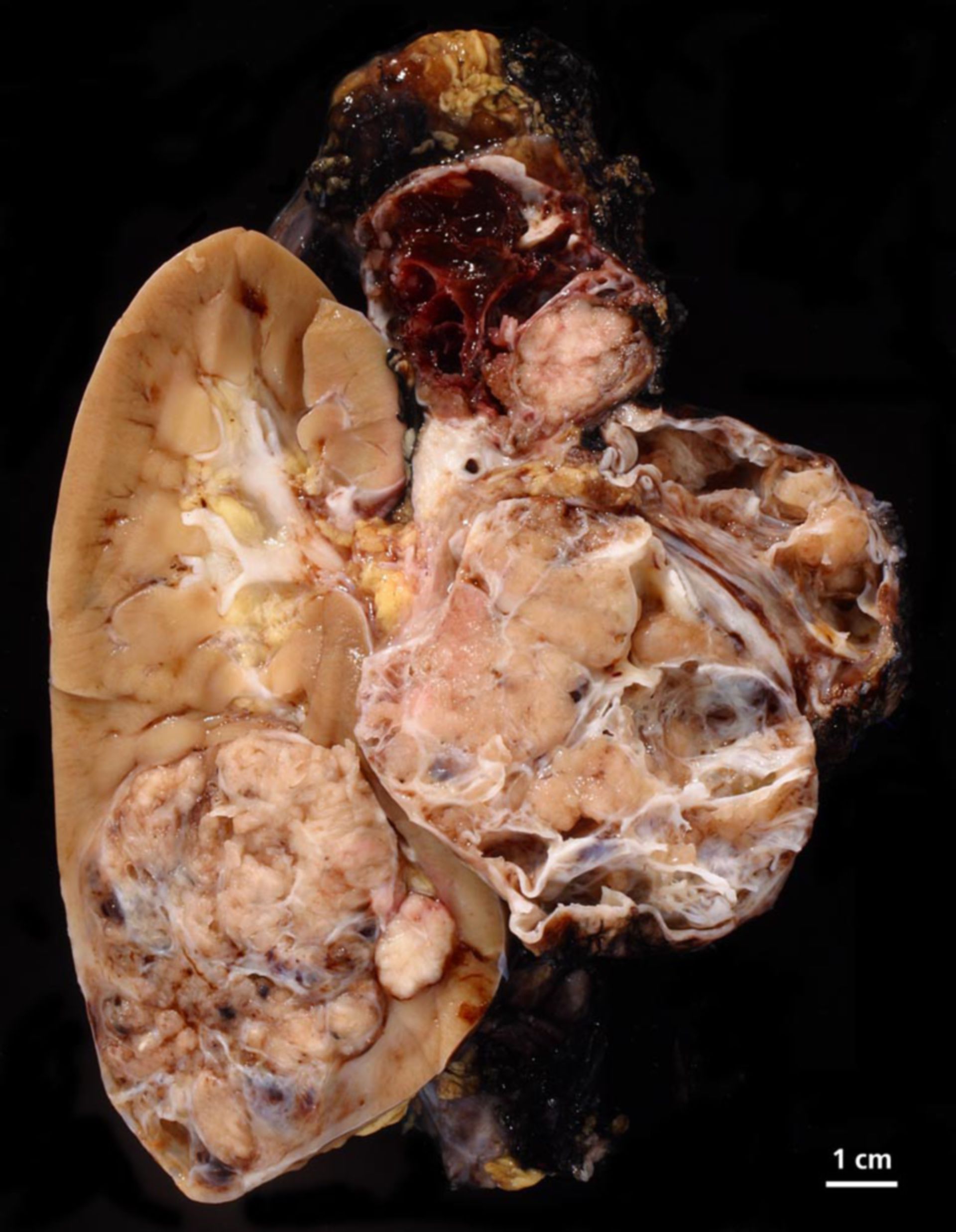
Description: On the section, in the lower end of the kidney, a localized tumor, polylobulated, max. diameter 6.7 cm. The tumor consists of necrosis and cysts. Two additional cystic tumor nodes (diameter 5 cm and 8 cm) with bleeding, next to the hilus. The kidney parenchyma with normal cortex-medulla-border and cortex up to 0.9 cm.
Additional findings: Immunohistological colorization for TFE-3, the translocation product of t(x;17) (p11.2;q25.3) or t(x;1) (p11.2;q21). The reaction was postive - clear nuclear colorization - which molecularly confirms the suspicion of a translocation carcinoma.
Clinically: 16-year old female patient with unclear retroperetonial tumor on the left side.
Commentary: Review: Clin Lab Med. 2005 Jun ;25(2):363-78. Translocation carcinomas of the kidney. Argani P, Ladanyi M.
Source: ©PathoPic