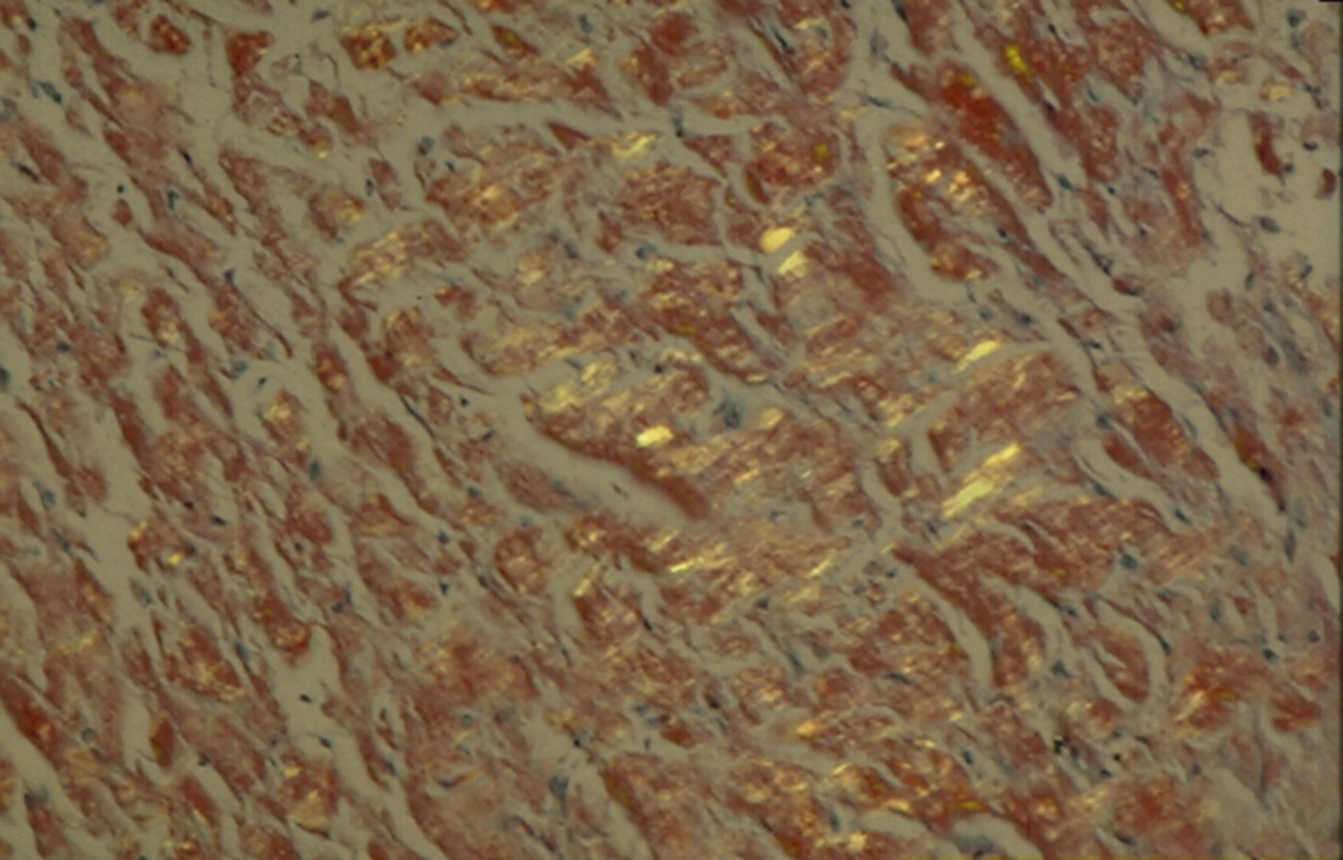
The biopsy material is stained with the congo red dye after histological preparation and after that, it is examined in immunohistochemistry. Amyloid binds Congo red and becomes visible under polarized light in the form of greenish luminous deposits in the intercellular space.