Mediastinal schwannomas are typically benign and asymptomatic, and generally present no immediate risks. We encountered a rare case of a giant benign posterior mediastinal schwannoma, complicated by life-threatening cardiac tamponade.
Case presentation
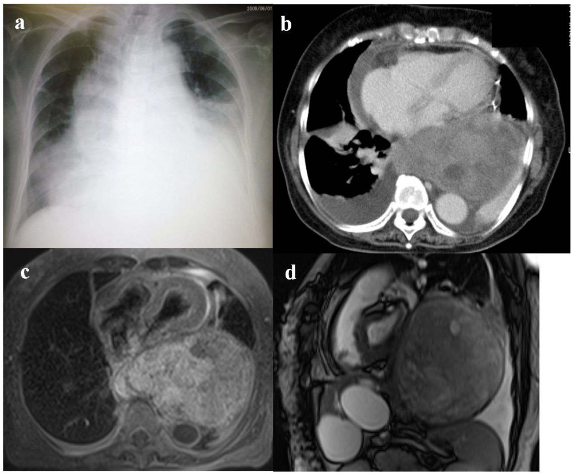
We report the case of a 72-year-old Japanese woman, who presented with cardiogenic shock. Computed tomography of the chest revealed a posterior mediastinal mass 150 cm in diameter, with pericardial effusion. The cardiac tamponade was treated with prompt pericardial fluid drainage. A biopsy was taken from the mass, and after histological examination, it was diagnosed as a benign schwannoma, a well-encapsulated non-infiltrating tumor, originating from the intrathoracic vagus nerve. It was successfully excised, restoring normal cardiac function.
Conclusion
Our case suggests that giant mediastinal schwannomas, although generally benign and asymptomatic, should be excised upon discovery to prevent the development of life-threatening cardiopulmonary complications.Lung imaging. (a) Chest radiograph obtained on the day of hospital admission showing bilateral pleural effusion, mediastinal widening and cardiac enlargement. (b) Contrast-enhanced chest computed tomography image (lung window) taken during pericardial drainage, showing a giant posterior mediastinal tumor, pericardial effusion and bilateral pleural effusion. (c) Transversal T1-weighted magnetic resonance imaging (MRI) scan of the chest taken after pericardial drainage, showing a giant encapsulating tumor in the posterior mediastinum compressing the heart. (d) Sagittal True SSFP (steady state free precession) MRI image, showing the tumor occupies most of left thoracic cavity.Source: Kato et al. Journal of Medical Case Reports 2011 5:61 doi:10.1186/1752-1947-5-61