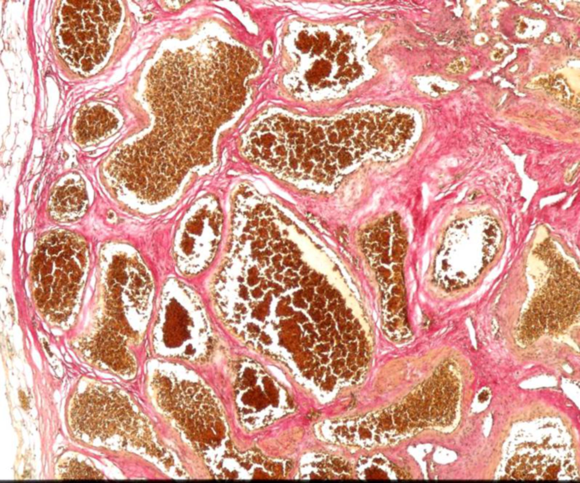
Histology of a cavernoma.
There are multiple pathological vessels visible, showing fibrinous thinned walls. EVG-stain.
Hemangiomas are tumors of the vessels. There are very common, especially in infants and toddlers.
In the cavernous hemangioma the malformations apper as red-blue spots, also called port wine spots.
They usually manifest on the skin but can also affect parenchymatous organs and the central nervous system. There is no capsula in the cavernous hemangioma. Under the microscope the blood-filled wide vessels with flat epithelium can be seen. Additionally there are single vessels, separated by connective tissue.
A complication of the cavernous hemangioma is the formation of a thrombus. In contrast to a capillary hemangioma, the tumor of the cavernous hemangioma does not spontaneously regress.
Source: Marvin 101 via wikipedia