CIL:44601 - http://www.cellimagelibrary.org/images/44601
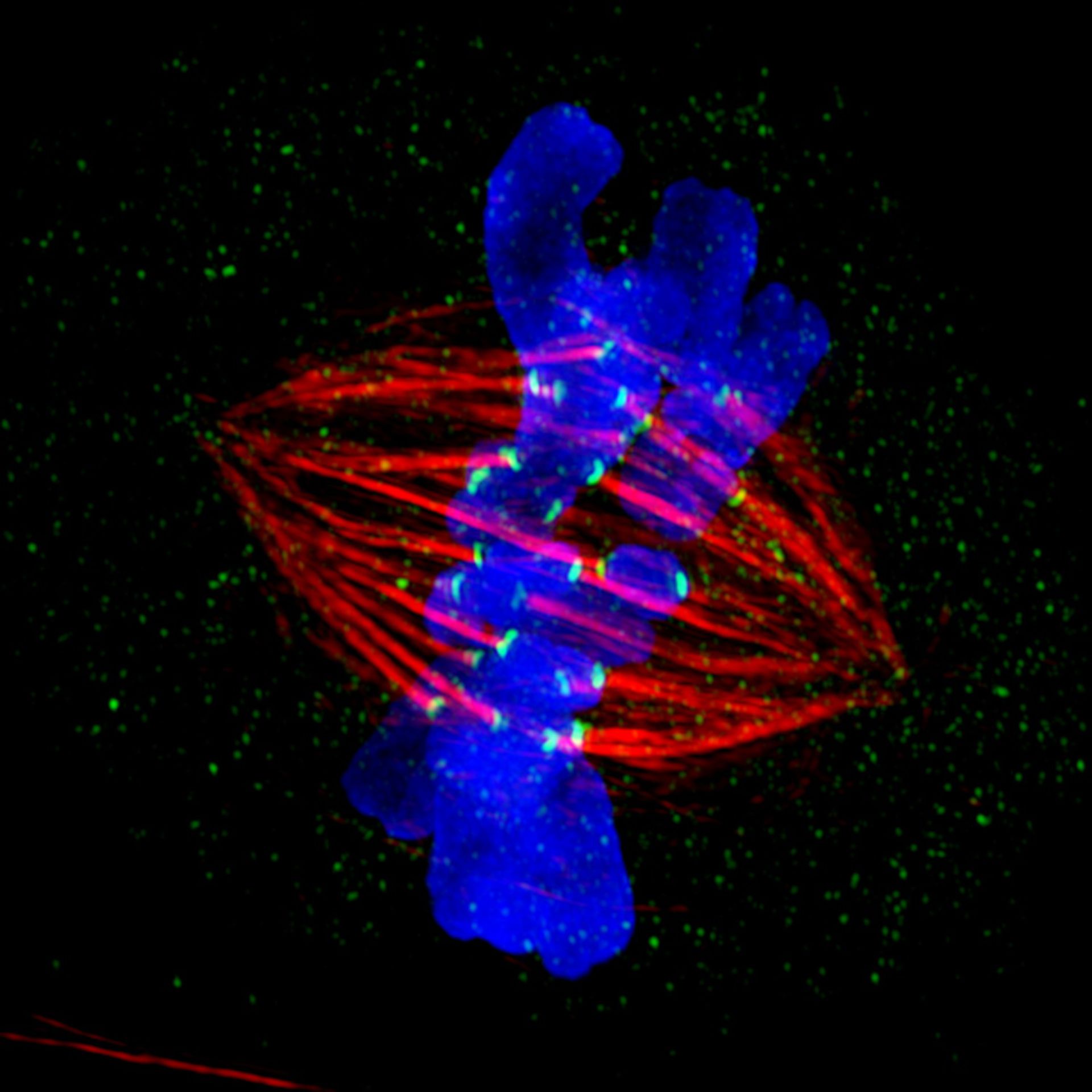
Description: Shown is a mitotic PtK2 cell at metaphase immunostained for microtubules (red) and kinetochores (green) with DNA stained blue. The image was obtained using structured illumination microscopy (SIM, Deltavision OMX system) which provides 'super-resolution' beyond the diffraction limit set by the wavelength of the illuminating light. The micrograph was the 2012 winner in the high- and super-resolution microscopy category of the GE Healthcare Life Sciences Imaging Contest, and featured in the NIGMS Biomedical Beat, the monthly digest of notable NIGMS-sponsored research.
Authors: Jane Stout and Claire Walczak
Licensing: Public Domain: This image is in the public domain and thus free of any copyright restrictions. However, as is the norm in scientific publishing and as a matter of courtesy, any user should credit the content provider for any public or private use of this image whenever possible.