CIL:39088 - http://www.cellimagelibrary.org/images/39088
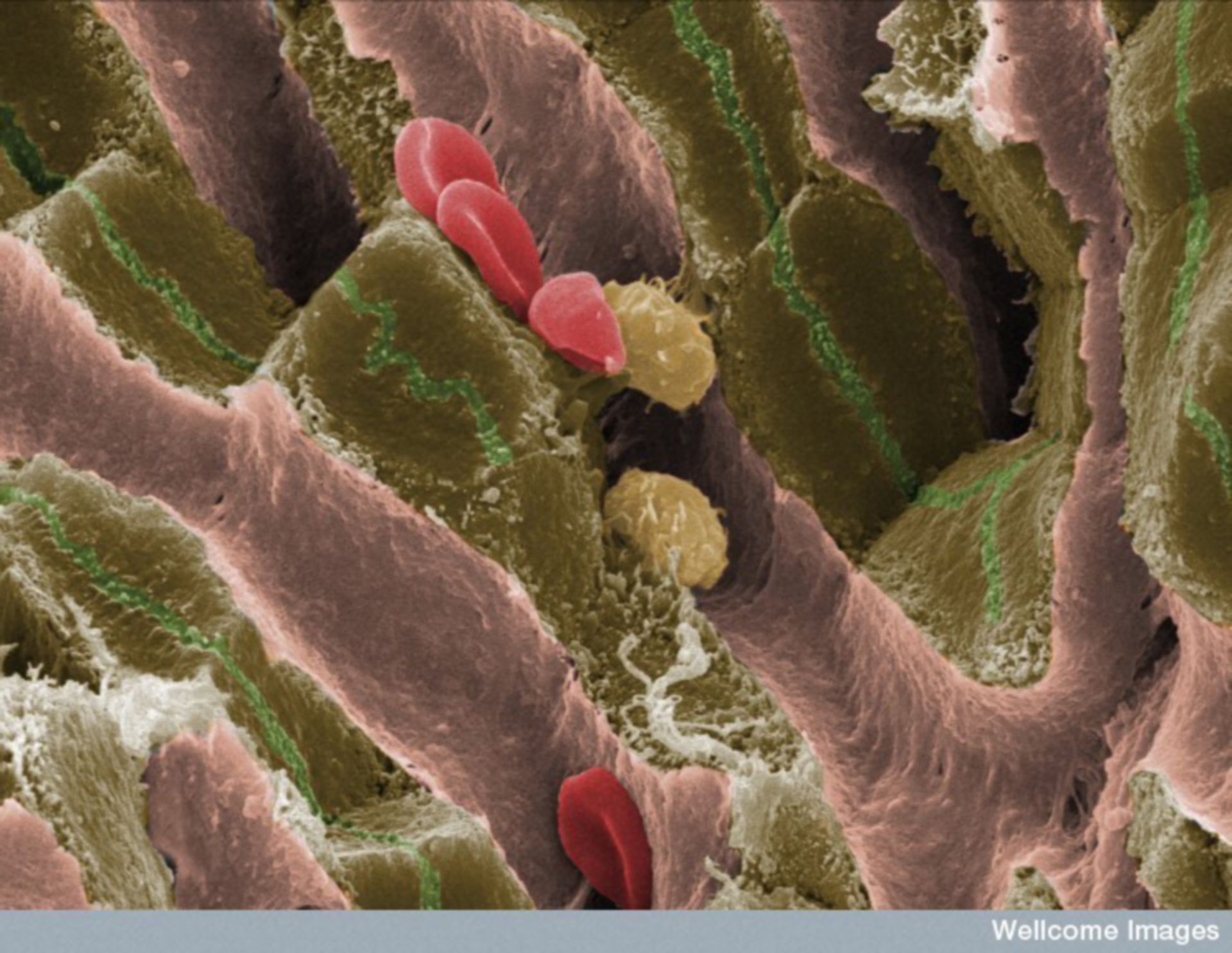
Description: This scanning electron micrograph shows the internal structure of liver tissue from an adult mouse. The sinusoids (vascular channels lined with endothelial cells) can be seen as pink structures running through the tissue. These contain red blood cells and Kupffer cells (specialized macrophages of the liver). Hepatocytes, shown in brown, are arranged in plates surrounding the sinusoids. Bile is secreted into the canaliculi, shown as green channels. These are dilated intercellular spaces between adjacent hepatocytes and bile flows through them en route to the small intestine.
Author: EM Unit, UCL Medical School, Royal Free Campus and Wellcome Images
Licensing: Attribution-NonCommercial-NoDerivs 2.0 UK: England & Wales (CC BY-NC-ND 2.0 UK)