Melanoma! Image of the Week - June 5, 2017
CIL:38978 - http://www.cellimagelibrary.org/images/38978
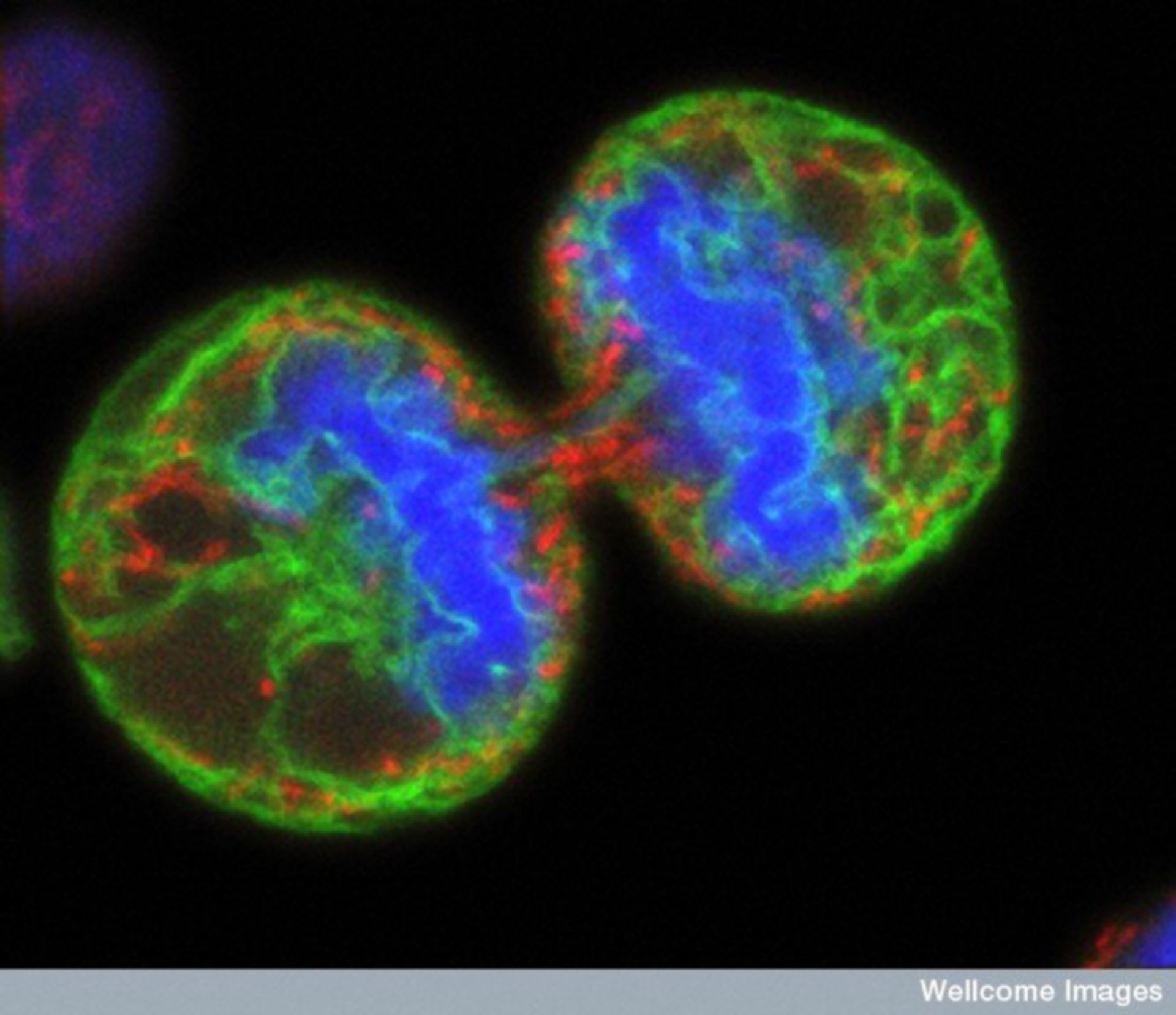
Description: Human melanoma cell undergoing cell division. The chromosomes (blue) have separated and the two daughter cells have almost split apart - only a small bridge of cytoplasm remains. The green staining labels the endoplasmic reticulum and the red labels the mitochondria. The image was produced on a confocal microscope; the ER and mitochondria are from a single optical section but the chromosomes are a 3D reconstruction from a series of sections.
Authors: Paul J. Smith and Rachel Errington
Licensing: Attribution-NonCommercial-NoDerivs 2.0 UK: England & Wales (CC BY-NC-ND 2.0 UK)