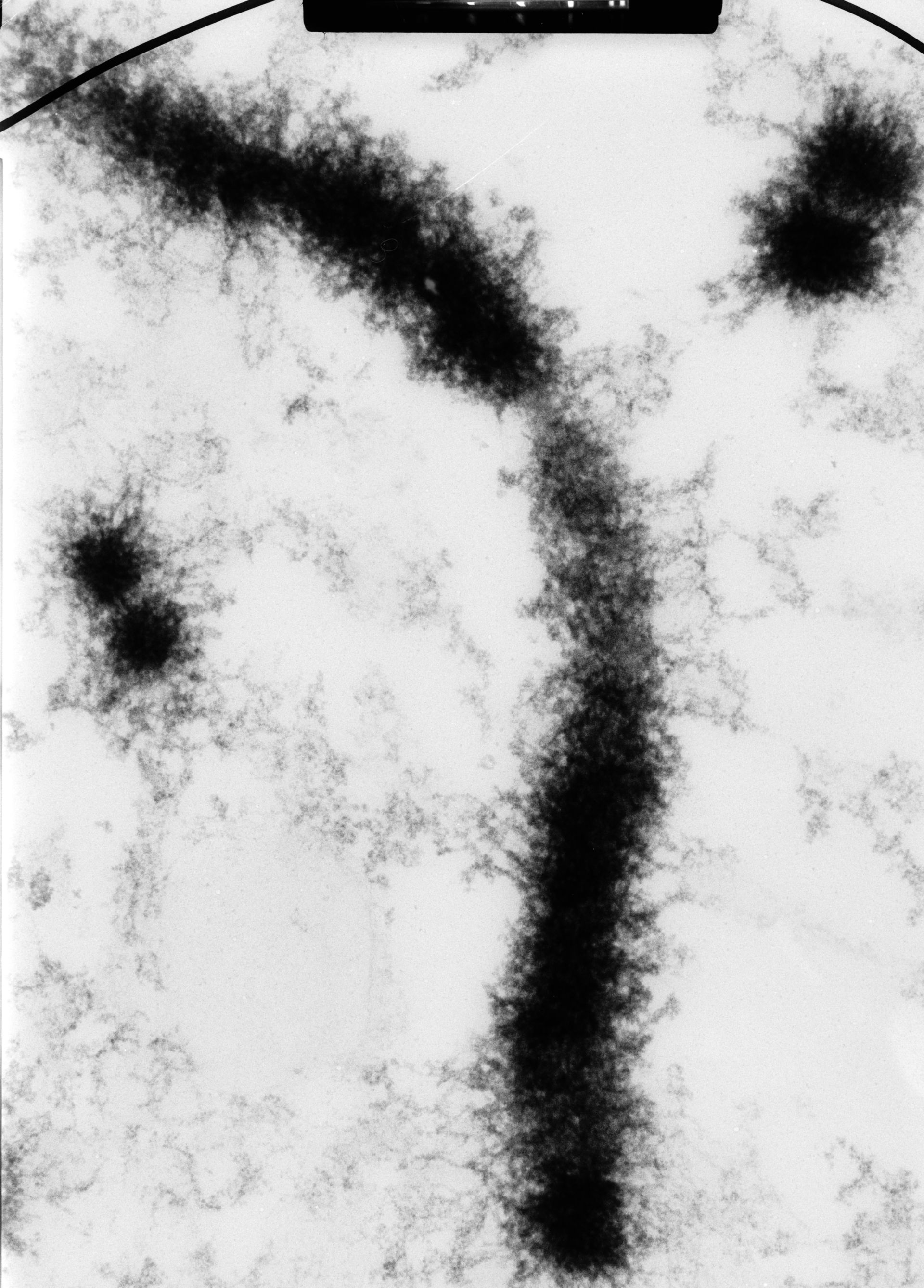
High voltage (1MeV) transmission electron microscopy image of a 0.5 micrometer section of a Chinese hamster ovary cell in metaphase containing a longitudinal section of a chromatid pair, and revealing the fiber-like structure. The image was taken with a specimen tilt of 45 degrees. Grouped with it is an image with a tilt of 55 degrees, providing a pair that affords an oblique stereo view of the chromosome.
Biological Process: Chromosome organization, Mitosis, Mitotic metaphase
Cells in metaphase were fixed, embedded in plastic, and 0.5 um sections cut and stained with uranyl acetate. See also: H. Ris 1981 Steroscopic electron microscopy of chromosomes. Meth Cell Biol 22:77-96 H. Ris 1978 Preparation of chromatin and chromosomes for electron microscopy. Meth Cell Biol 18:220-246.
Author: Hans Ris
Source: The Cell: An Image Library