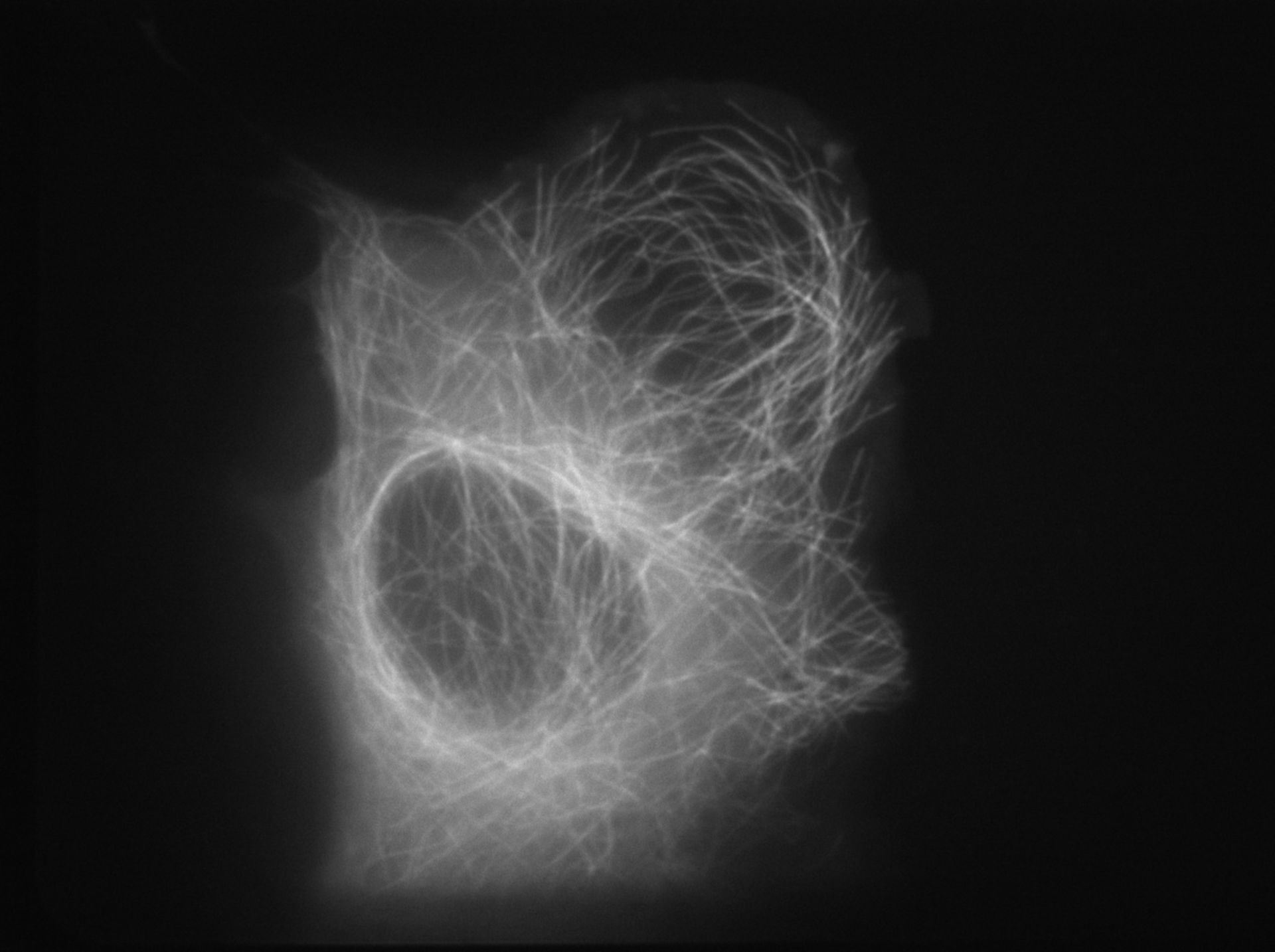
Microtubule dynamics in a U2OS (human osteosarcoma) cell. Cells were injected with Xrhodamine-tubulin and this time-lapse image series was collected on a spinning disk confocal microscope on a Nikon Ti200 using a 100X 1.4 NA Plan Apo objective with a 605x52 EM filter for 568 nm laser excitation. Images were collected with a Coolsnap HQ2 (Photometrics) with 700 ms exposure.
Biological Process: Microtubule cytoskeleton organization
Author: Clare M. Waterman
Source: The Cell: An Image Library