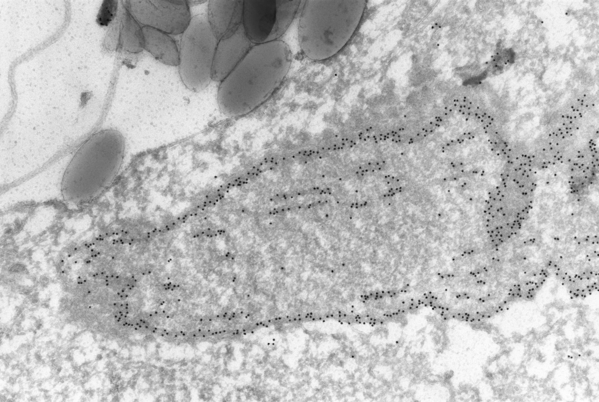
High resolution view during division showing the micronucleus contains internal microtubules as well as a layer of b-tubulin, not in microtubular form, on or just inside the nuclear envelope. The section is immunogold labeled with anti-b-tubulin. TEM taken on 7/8/96 by R. Allen with Zeiss 10A operating at 60kV. Neg. 19,800X. Cells were lightly fixed with 0.25% glutaraldehyde and infiltrated with 2.3M sucrose before being frozen in liquid nitrogen and thin sectioned at a temperature of –100?C at approximately 75nm thickness. Frozen sections from these preparations were then thawed, washed, and exposed to a monoclonal primary antibody that was raised in mice or rabbit/goat and to colloidal gold-complexed goat-anti-mouse/rabbit secondary antibodies. Further details of preparation are detailed in Methods Cell Biol. 2010;96:143-73. The raw film was scanned with a Nikon Coolscan 9000ED. This image is best used for quantitative analysis. Additional information available at (http://www5.pbrc.hawaii.edu/allen/).
Biological Process: Nuclear division, Microtubule-based process, Microtubule-based process
Author: Richard Allen (University of Hawaii)
Source: The Cell: An Image Library