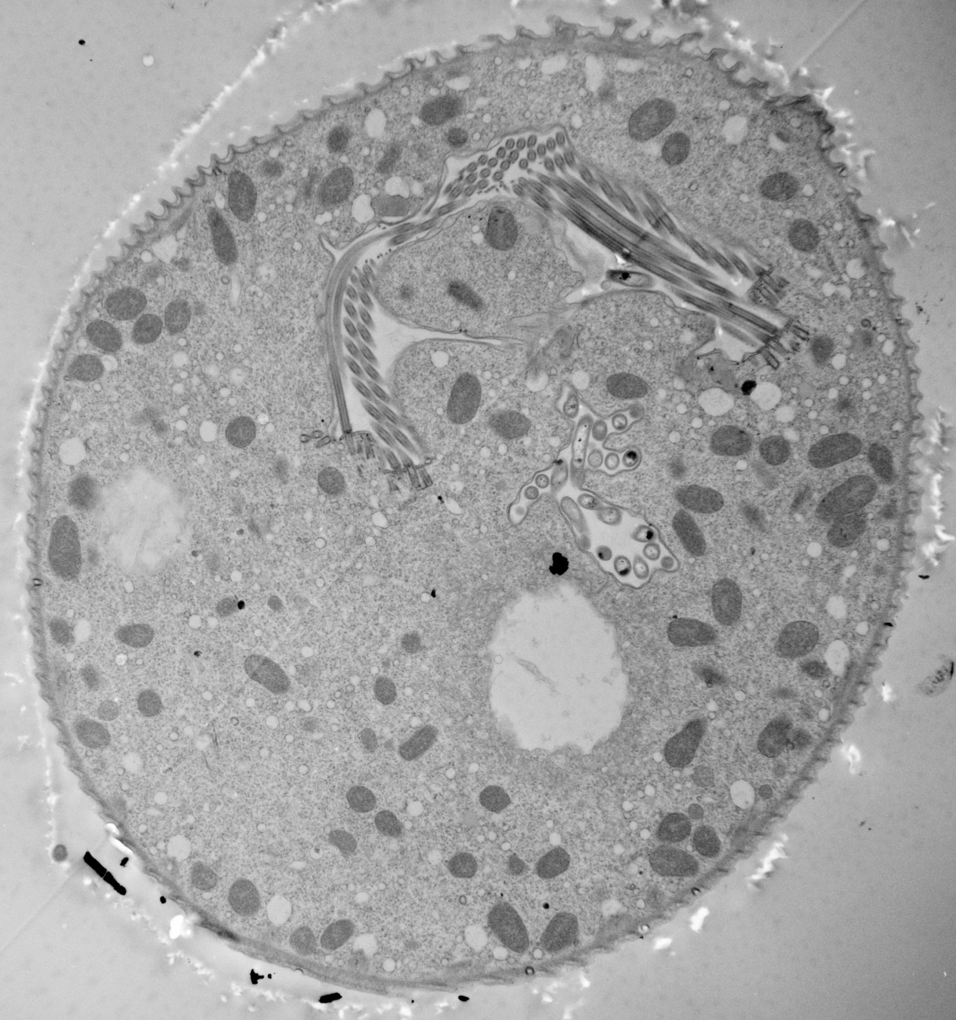
The 11 micrographs of the group are views of a serially-sectioned contracted Vorticella convallaria cell that show the main features of this cell. This figure shows the edge of last food vacuole in series; contractile vacuole; bacteria-filled chamber; peristome open to the perioral chamber that opens to the cell’s exterior. TEM taken on 4/1/71 By R. Allen with Hitachi HU11A operating at 75kV. Neg. 2,650X. The raw negative was scanned with an Epson Perfection V750 Pro and this high resolution image is best used for quantitative analysis. Additional information available at (http://www5.pbrc.hawaii.edu/allen/).
Biological Process: Digestive system process, Cytoplasm organization, Cortical cytoskeleton organization, Oral apparatus organization, Contractile vacuole organization
Standard glutaraldehyde fixation followed by osmium tetroxide, dehydrated in alcohol and embedded in an epoxy resin. Microtome sections prepared at approximately 75nm thickness. Additional information available at (http://www5.pbrc.hawaii.edu/allen/).
Author: Richard Allen (University of Hawaii)
Source: The Cell: An Image Library