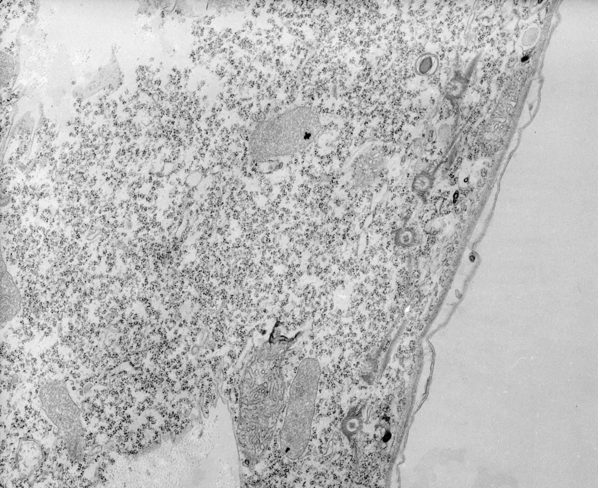
During division new basal bodies develop accessory fibers and begin to move apart to take up a position equidistant between the older complexes. The new kinetodesmal fiber seems to be associated with the prexisting kinetodesmal fiber during separation. TEM taken on 8/15/67 by R. Allen with Philips 200 operating at 60kV. Neg. 12,400X. The raw negative was scanned with an Epson Perfection V750 Pro and this high resolution image is best used for quantitative analysis. Additional information available at (http://www5.pbrc.hawaii.edu/allen/).
Biological Process: Microtubule basal body duplication
Standard glutaraldehyde fixation followed by osmium tetroxide, dehydrated in alcohol and embedded in an epoxy resin. Microtome sections prepared at approximately 75nm thickness. Additional information available at (http://www5.pbrc.hawaii.edu/allen/).
Author: Richard Allen (University of Hawaii)
Source: The Cell: An Image Library