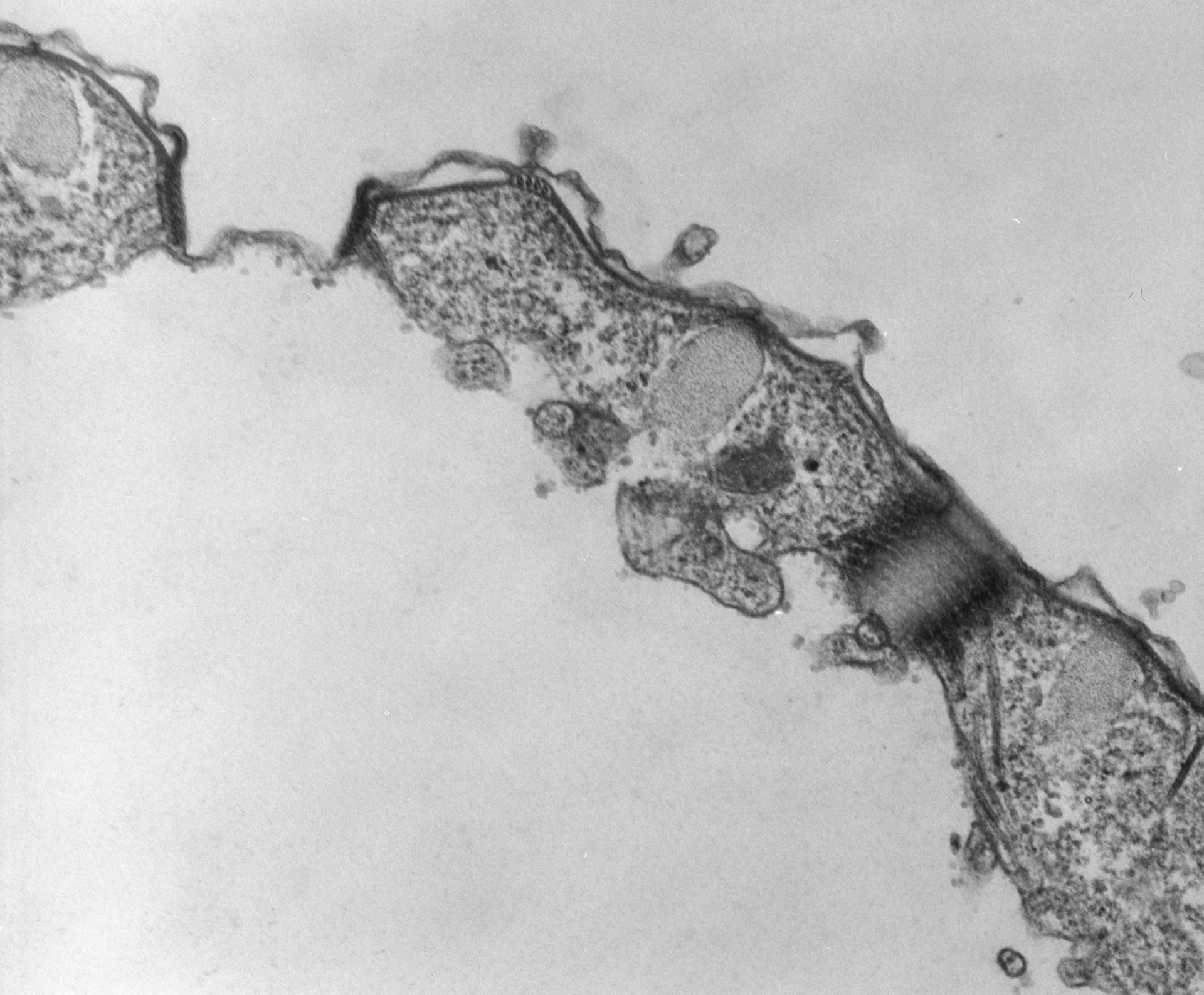
The two CV pores of a cell. An impression of the helically wound microtubules are detectable in the right pore. TEM taken on 4/21/78 by R. Allen with Hitachi HU11A operating at 60kV. Neg. 21,750X. The raw negative was scanned with an Epson Perfection V750 Pro and this high resolution image is best used for quantitative analysis. Additional information available at (http://www5.pbrc.hawaii.edu/allen/).
Biological Process: Contractile vacuole organization
Standard glutaraldehyde fixation followed by osmium tetroxide, dehydrated in alcohol and embedded in an epoxy resin. Microtome sections prepared at approximately 75nm thickness. Additional information available at (http://www5.pbrc.hawaii.edu/allen/).
Author: Richard Allen (University of Hawaii)
Source: The Cell: An Image Library