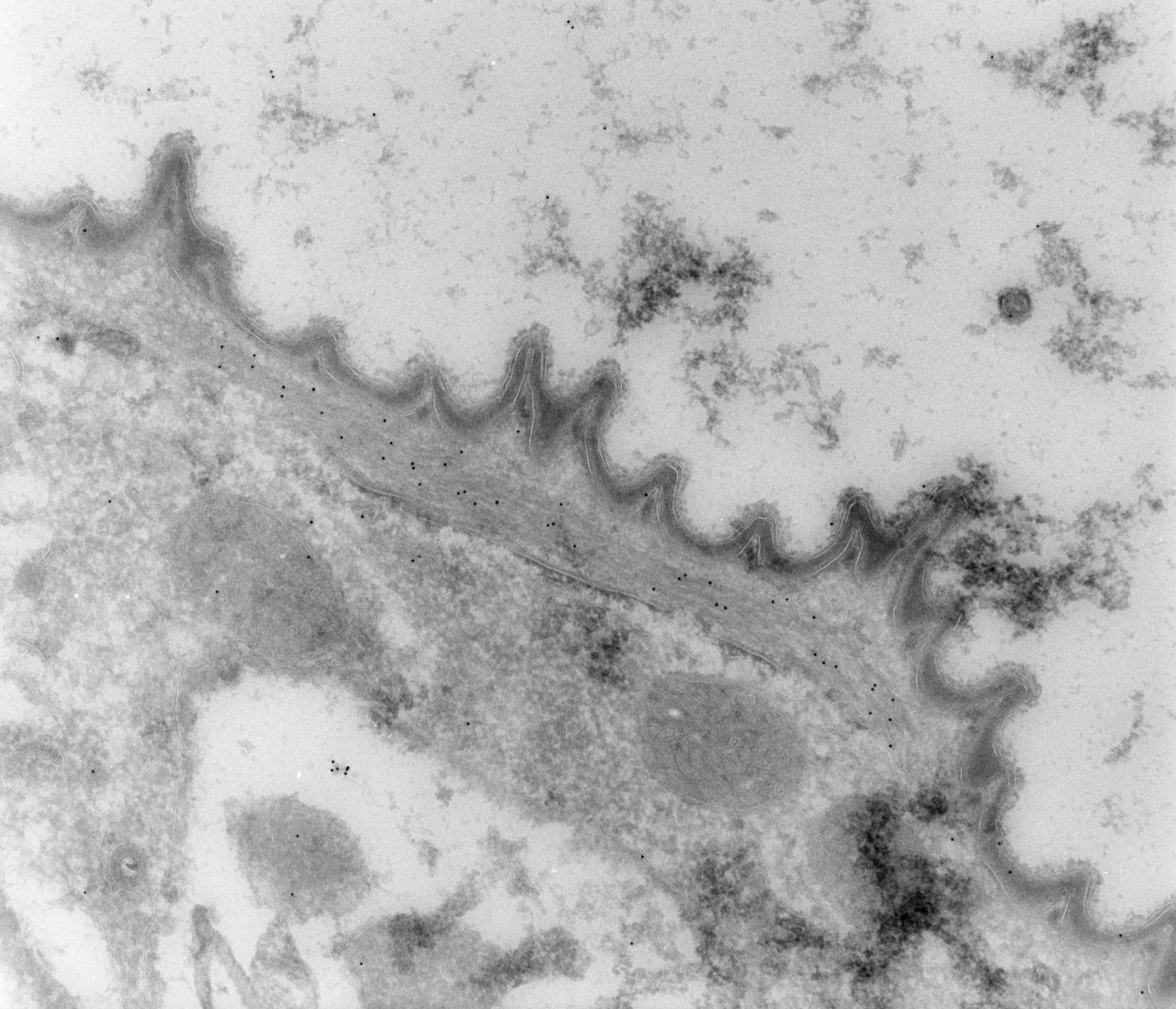
A thin cryosection of Vorticella exposed to a rabbit polyclonal antibody to alpha-centrin and then to gold conjugated to anti-rabbit IgG. The gold indicates that the myoneme contains alpha-centrin. The plasma membrane, outer alveolar membrane and inner alveolar membrane all appear as transparent tracks but the lumen of the alveolus appears electron opaque. Thickenings of the ER lying against the myoneme correspond to linkage complexes. TEM taken on 8/18/94 by D. Kunkel with Zeiss 10A operating at 80kV. Neg. 19,800X. The raw negative was scanned with an Epson Perfection V750 Pro and this high resolution image is best used for quantitative analysis. Additional information available at (http://www5.pbrc.hawaii.edu/allen/).
Biological Process: Regulation of myofibril size, Plasma membrane organization, Centrin localization
Cells were lightly fixed with 0.25% glutaraldehyde and infiltrated with 2.3M sucrose before being frozen in liquid nitrogen and thin sectioned at a temperature of –100?C at approximately 75nm thickness. Frozen sections from these preparations were then thawed, washed, and exposed to a monoclonal primary antibody that was raised in mice or rabbit/goat and to colloidal gold-complexed goat-anti-mouse/rabbit secondary antibodies. Further details of preparation are detailed in Methods Cell Biol. 2010;96:143-73.
Author: Richard Allen (University of Hawaii)
Source: The Cell: An Image Library