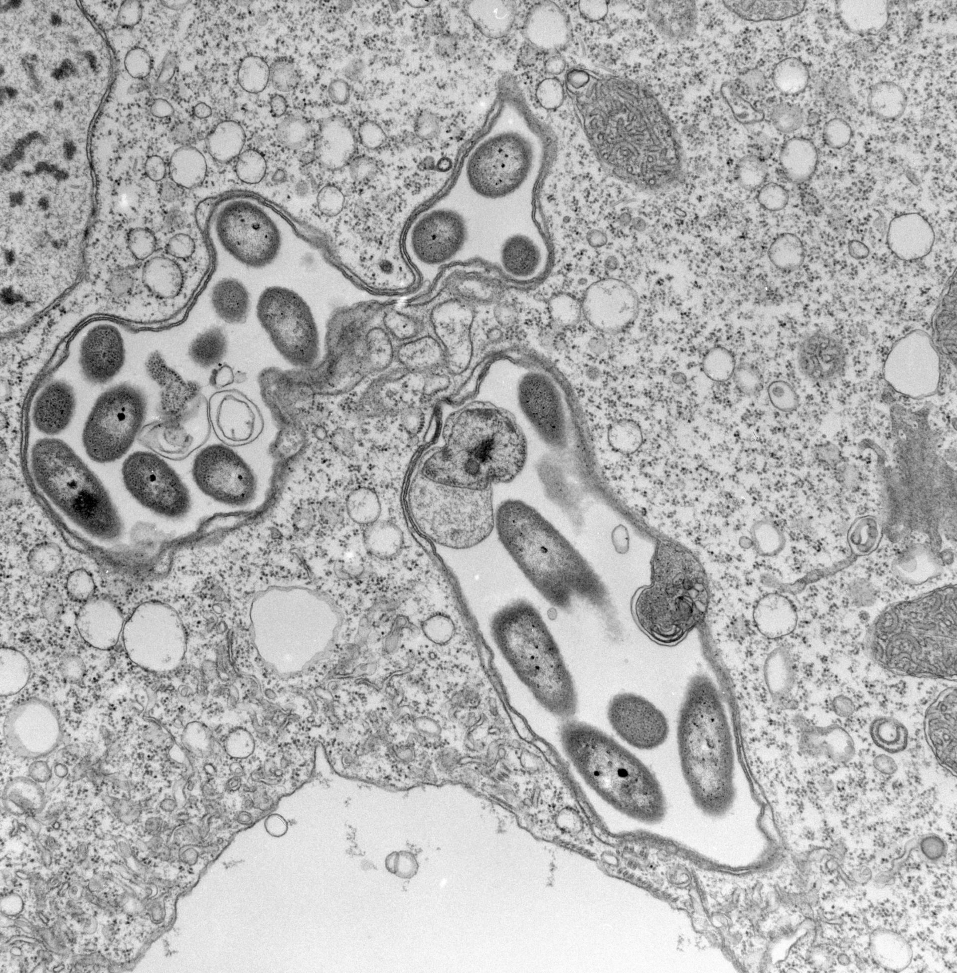
The contractile vacuole of V. convallaria empties into a convoluted chamber. This chamber is covered by the alveoli and plasma membrane of the pellicle and is open to the cell’s surface via the peristome which is in turn connected to the perioral opening in the contracted cell. This preCV chamber apparently harbors (cultures?) its own flora of bacteria. Ribbons of microtubules link the CV pore to the CV. TEM taken on 3/29/71 by R. Allen with Hitachi HU11A operating at 75kV. Neg. 11,250X. The raw negative was scanned with an Epson Perfection V750 Pro and this high resolution image is best used for quantitative analysis. Additional information available at (http://www5.pbrc.hawaii.edu/allen/).
Biological Process: Contractile vacuole organization, Microtubule-based process, Macronucleus organization
Standard glutaraldehyde fixation followed by osmium tetroxide, dehydrated in alcohol and embedded in an epoxy resin. Microtome sections prepared at approximately 75nm thickness. Additional information available at (http://www5.pbrc.hawaii.edu/allen/).
Author: Richard Allen (University of Hawaii)
Source: The Cell: An Image Library