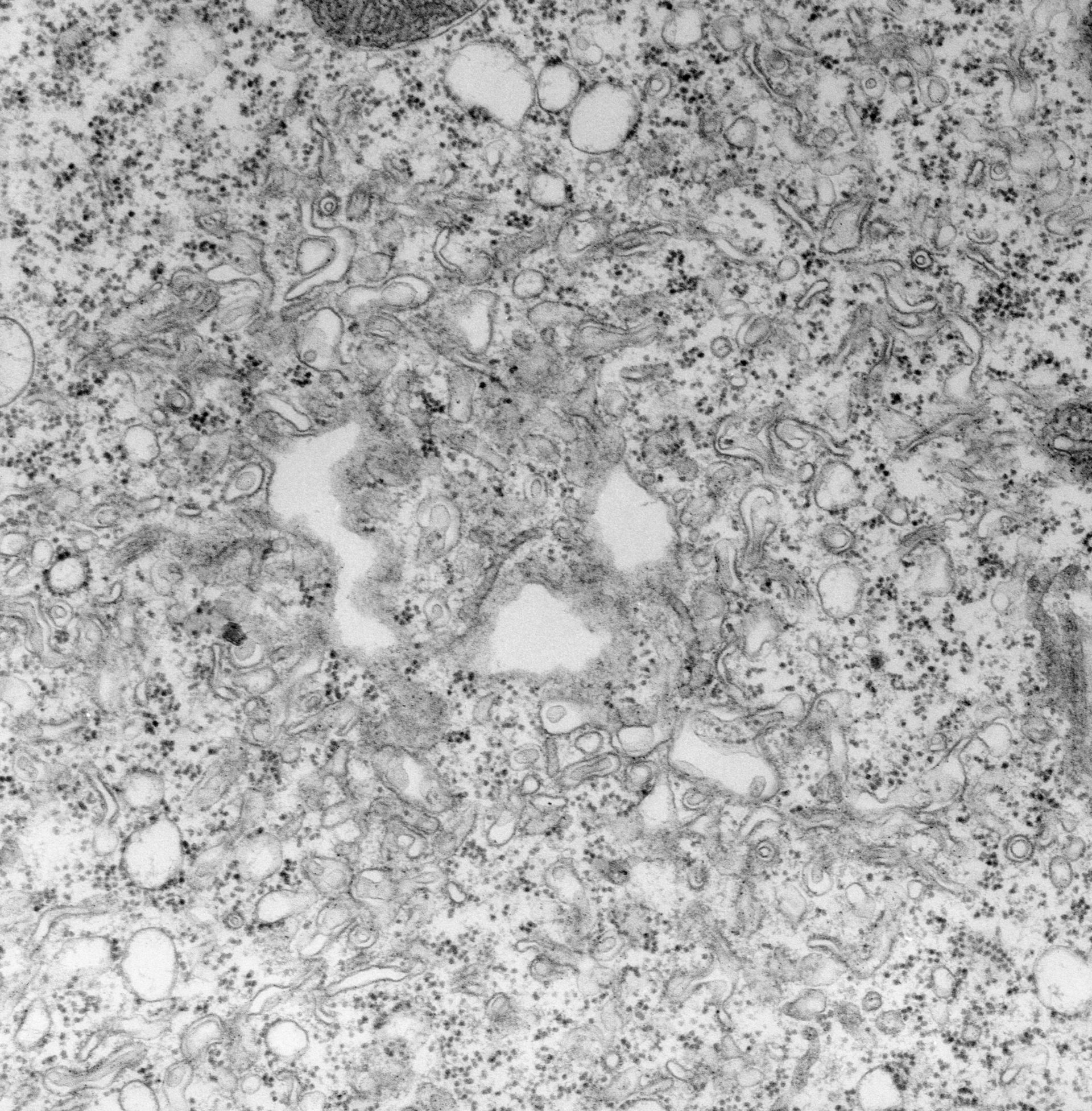
The contractile vacuole pore is supported on its cytosolic side by a coil of microtubules and other microtubules extend from the pore to the CV membrane. The plasma membrane and CV membrane cover the bottom of the pore. Bacteria reside in the chamber. TEM taken on 2/14/72 by R. Allen with Hitachi HU11A operating at 75kV. Neg. 19,500X. The raw negative was scanned with an Epson Perfection V750 Pro and this high resolution image is best used for quantitative analysis. Additional information available at (http://www5.pbrc.hawaii.edu/allen/).
Biological Process: Contractile vacuole organization, Plasma membrane organization, Microtubule-based process, Detection of symbiont
Standard glutaraldehyde fixation followed by osmium tetroxide, dehydrated in alcohol and embedded in an epoxy resin. Microtome sections prepared at approximately 75nm thickness. Additional information available at (http://www5.pbrc.hawaii.edu/allen/).
Author: Richard Allen (University of Hawaii)
Source: The Cell: An Image Library