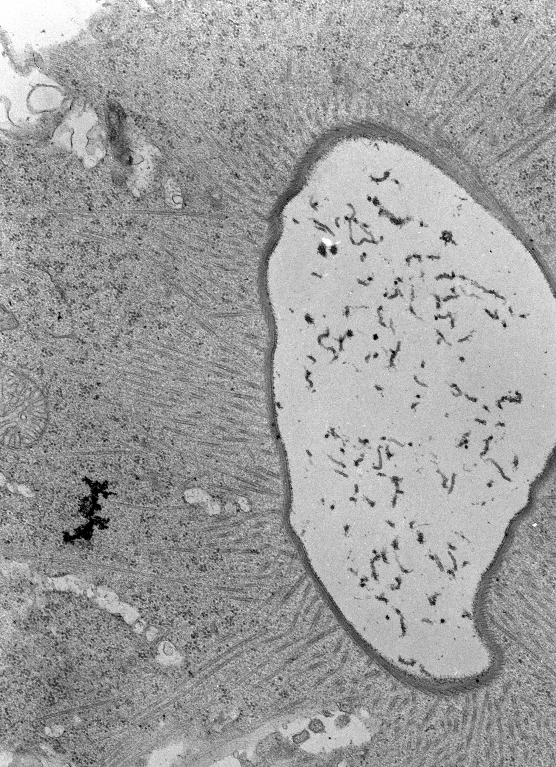
Detail of CV pore. Microtubules encircle the pore wall and other microtubules radiate out from the pore toward the CV membrane. TEM taken on 3/27/69 by R. Allen with Philips 300 operating at 60kV. Neg. 14,800X. The raw negative was scanned with an Epson Perfection V750 Pro and this high resolution image is best used for quantitative analysis. Additional information available at (http://www5.pbrc.hawaii.edu/allen/).
Biological Process: Contractile vacuole pore organization, Microtubule cytoskeleton organization, Cytoplasm organization
Standard glutaraldehyde fixation followed by osmium tetroxide, dehydrated in alcohol and embedded in an epoxy resin. Microtome sections prepared at approximately 75nm.
Author: Richard Allen
Source: The Cell: An Image Library