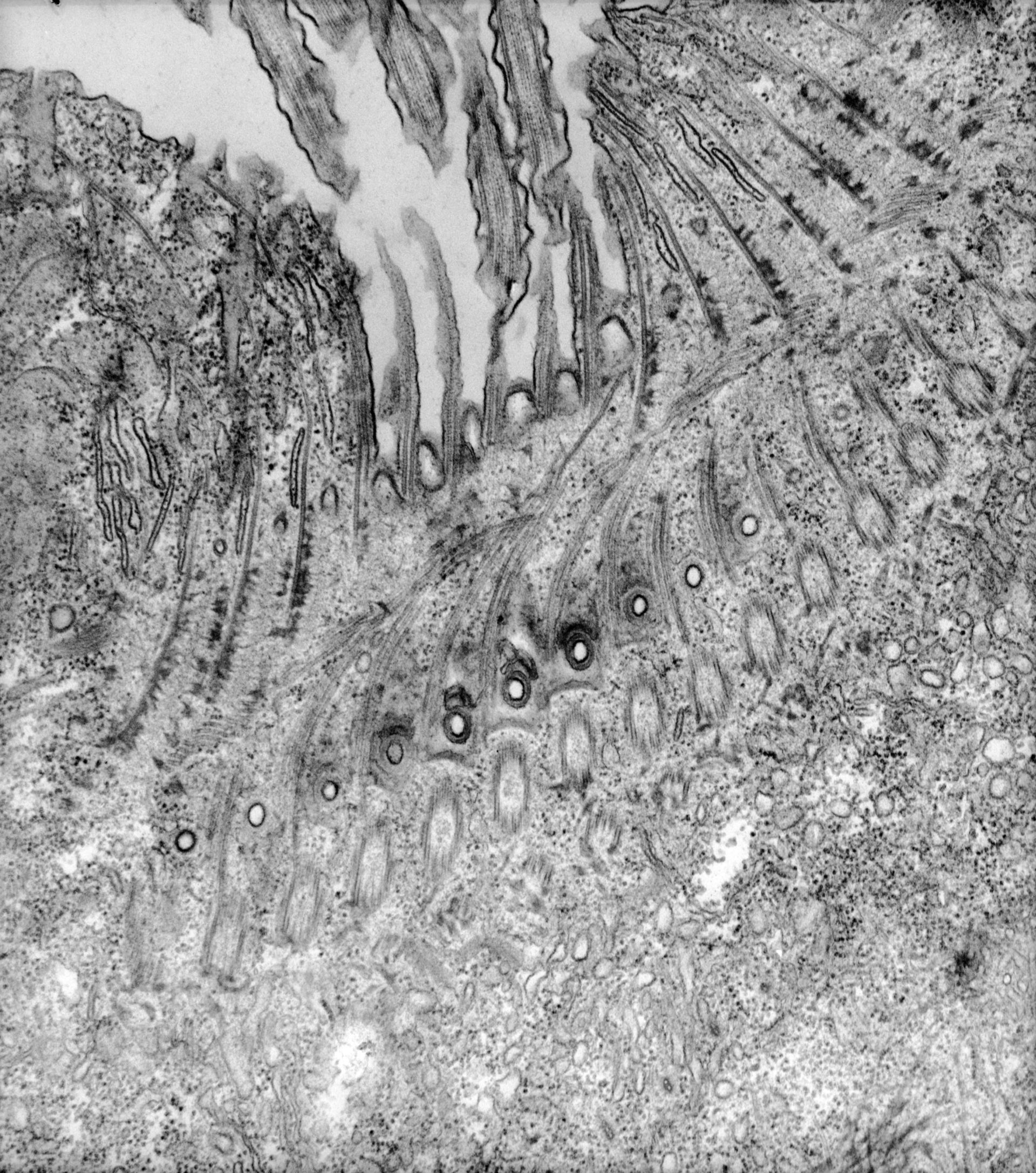
Details of the origins of the cytopharyngeal ribbons and the quadrulus associated ribbons. At first view the cytopharyngeal ribbons look like they might arise next to basal bodies but it is clear that the cytopharyngeal ribbons arise only from the filamentous reticulum. The quadrulus associated ribbons appear to be analogous to the postciliary microtubules of the somatic ciliature that they curve posteriorly from the quadrulus. However quadrulus associated ribbons do pass very close or even touch the ends of the cytopharyngeal ribbons without becoming part of these latter ribbons. Discoidal vesicles bind to the anterior sides (observer’s left) of the cytopharyngeal ribbons and the smaller set of ribbons lies at its posterior side against the cytopharyngeal membrane. The tubular fingers of the alveoli pass between the cytopharyngeal ribbons next to the nodes of the filamentous reticulum from which the cytopharyngeal ribbons originate. Finally a row of parasomal sacs is seen along the dorsal edge of the quadrulus from which coated vesicles form. TEM taken on 5/23/73 by R. Wolf with Hitachi HU11A operating at 75kV. Neg. 16,250X.
Biological Process: Oral apparatus organization
Standard glutaraldehyde fixation followed by osmium tetroxide, dehydrated in alcohol and embedded in an epoxy resin. Microtome sections prepared at approximately 75nm thickness. The raw negative was scanned with an Epson Perfection V750 Pro and this high resolution image is best used for quantitative analysis. Additional information available at (http://www5.pbrc.hawaii.edu/allen/).
Author: R. Wolf
Source: The Cell: An Image Library