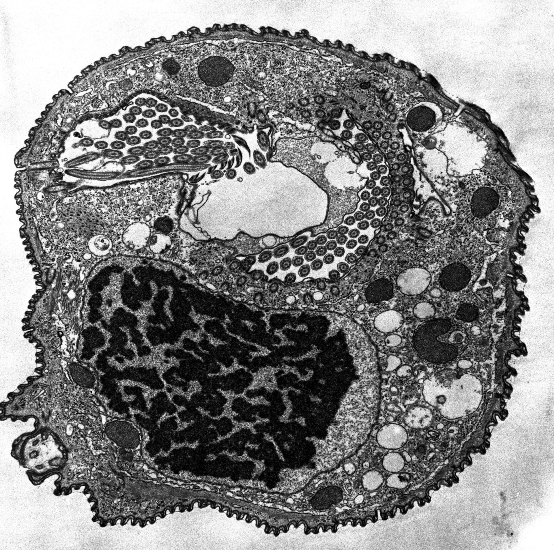
The peristome is covered on one side by the oral cilia and on the opposite by the ribbed wall. Lamellae from the ribbed wall extend into the nascent food vacuole forming region, the cytopharynx. This cell was not feeding and had no food vacuoles. TEM taken on 6/10/69 by R. Allen with Philips 300 operating at 60kV. Neg. 5,000X.
Biological Process: Macronucleus organization, Cortical cytoskeleton organization, Cytoplasm organization, Oral apparatus organization
Standard glutaraldehyde fixation followed by osmium tetroxide, dehydrated in alcohol and embedded in an epoxy resin. Microtome sections prepared at approximately 75nm thickness. The raw negative was scanned with an Epson Perfection V750 Pro and this high resolution image is best used for quantitative analysis. Additional information available at (http://www5.pbrc.hawaii.edu/allen/).
Author: Richard Allen
Source: The Cell: An Image Library