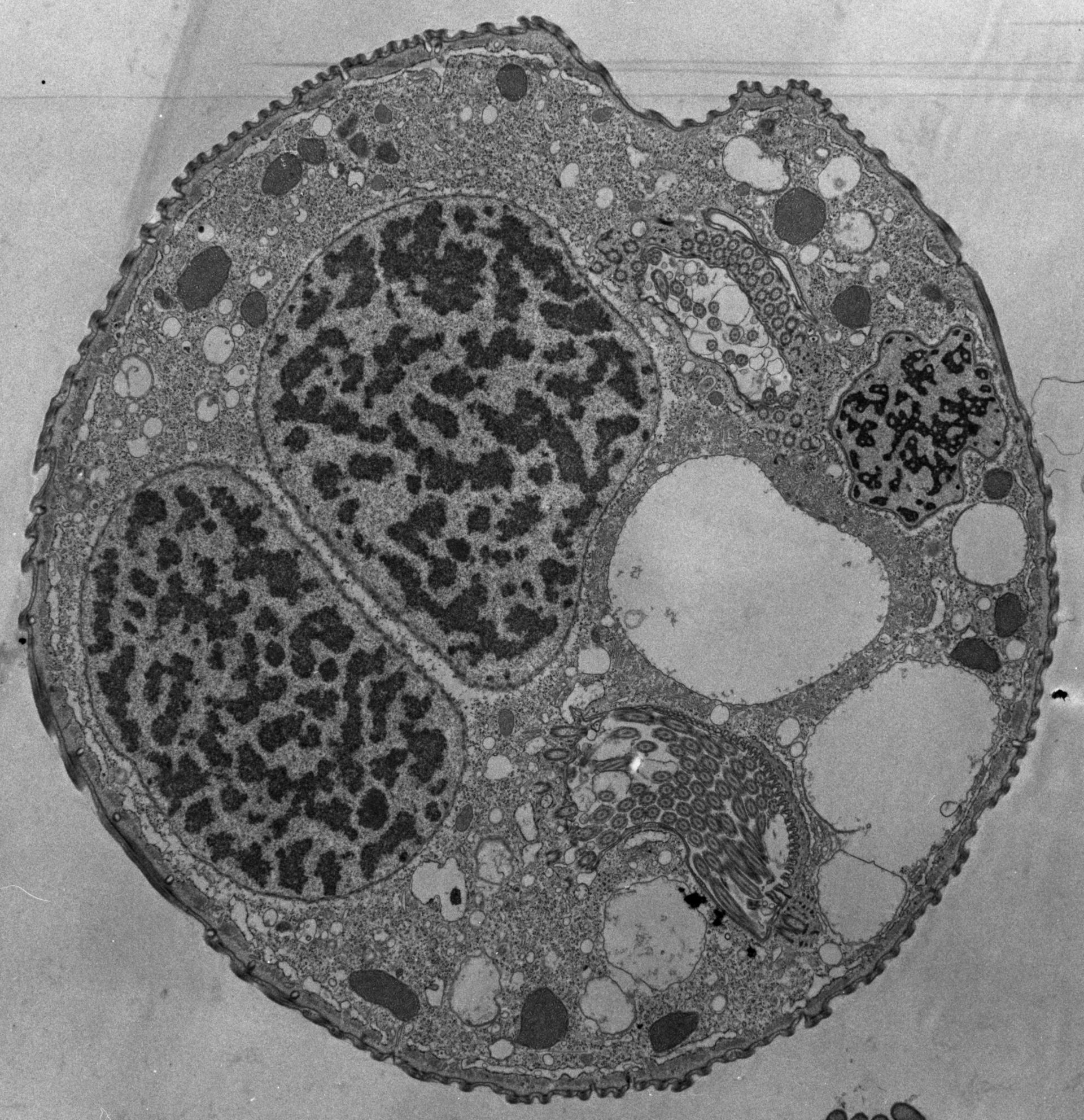
This section is cut at a level where the macronucleus is folded so it appears to be in 2 parts and where the micronucleus is visible. Contractile vacuole, perioral kinetids, and the peristome is evident. TEM taken on 6/10/69 by R. Allen with Philips 300 operating at 60kV. Neg. 3,570X.
Biological Process: Macronucleus organization, Cortical cytoskeleton organization, Cytoplasm organization, Contractile vacuole organization, Micronucleus organization
Standard glutaraldehyde fixation followed by osmium tetroxide, dehydrated in alcohol and embedded in an epoxy resin. Microtome sections prepared at approximately 75nm thickness. The raw negative was scanned with an Epson Perfection V750 Pro and this high resolution image is best used for quantitative analysis. Additional information available at (http://www5.pbrc.hawaii.edu/allen/).
Author: Richard Allen
Source: The Cell: An Image Library