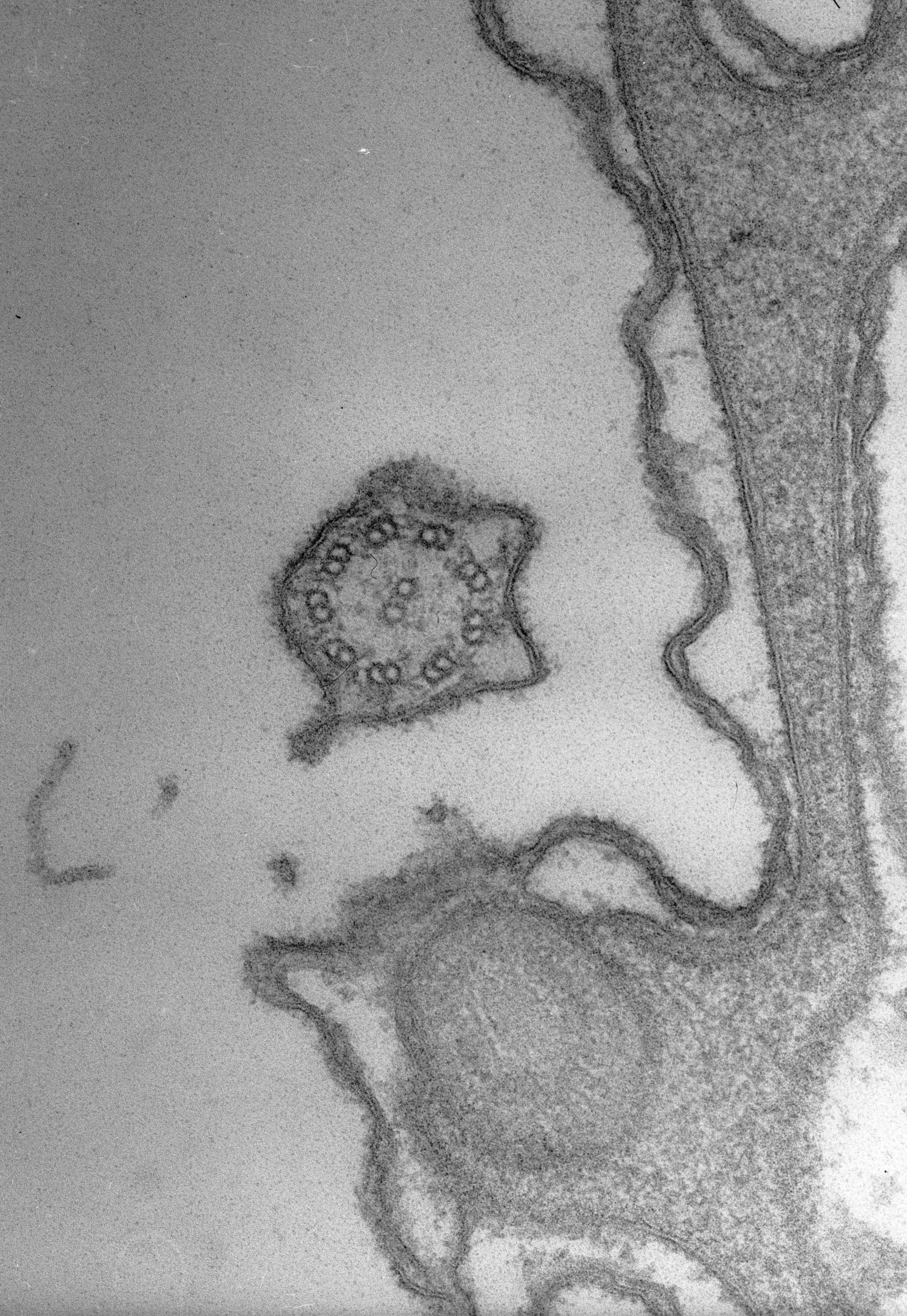
A higher magnification view that shows the bilayer nature of the ciliary membrane, plasma membrane and the outer and inner (alveolar sac membranes of the alveoli. The 9-doublet + 2-singlet microtubular axonemal system of the cilium is seen in cross section. Two dynein arms extend from each doublet. Cacodylate buffered fixative. TEM taken on 9/27/68 by R. Allen with Philips 300 operating at 60kV. Neg. 60,400X.
Biological Process: Cilium organization, Cortical cytoskeleton organization
Standard glutaraldehyde fixation followed by osmium tetroxide, dehydrated in alcohol and embedded in an epoxy resin. Microtome sections prepared at approximately 75nm thickness. The raw negative was scanned with an Epson Perfection V750 Pro and this high resolution image is best used for quantitative analysis. Additional information available at (http://www5.pbrc.hawaii.edu/allen/).
Author: Richard Allen (University of Hawaii)
Source: The Cell: An Image Library