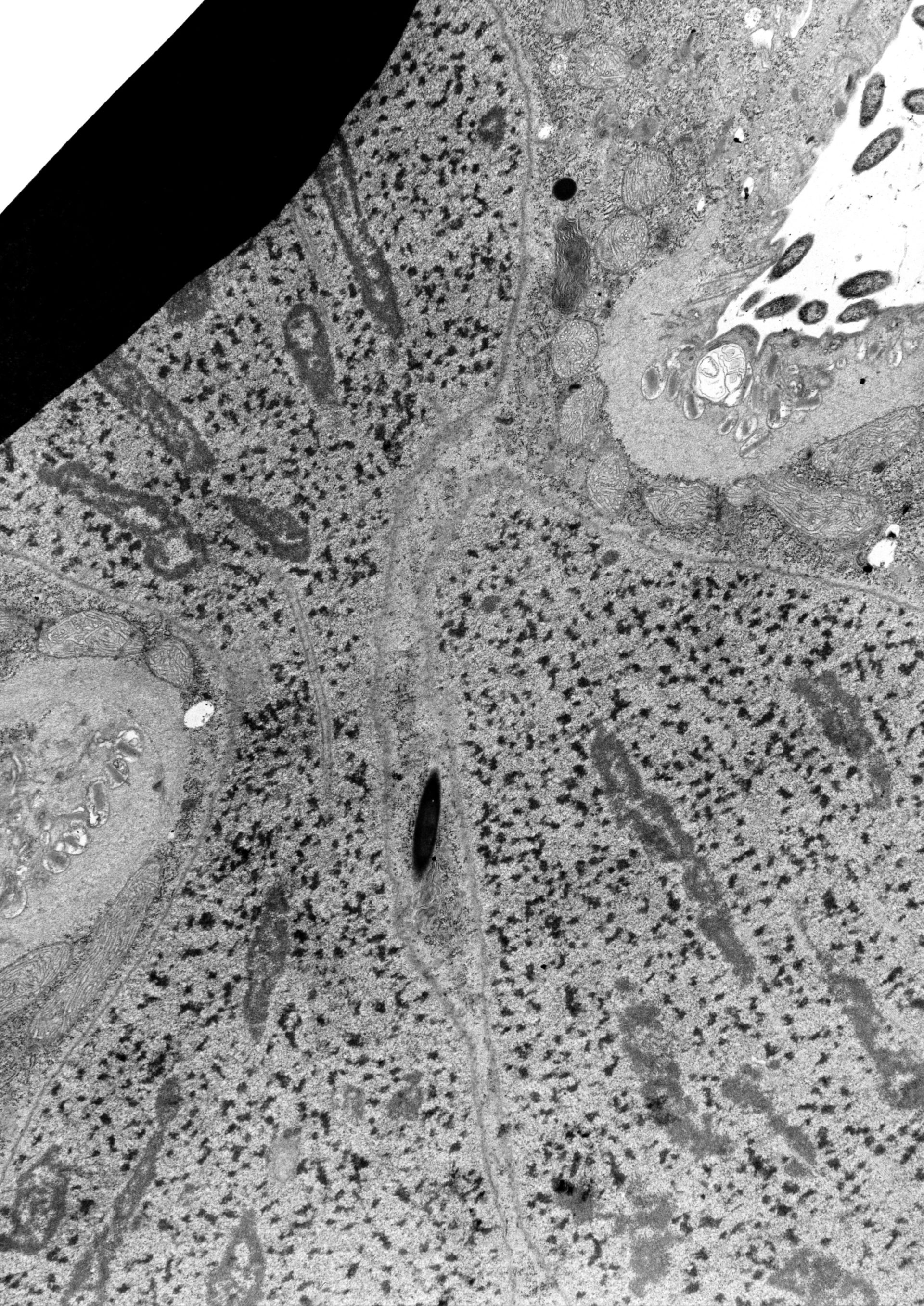
The division furrow of a dividing Didinium. A thick layer of epiplasm coated on the cytosolic side by mitochondria forms the constricting ring in the furrow. As usual mucocysts lie between the epiplasm and the pellicular membranes. The new cytopharynx in the opisthe already has lamellae and the alveolus seems to be absent from this region. The macronucleus is constricted in the furrow and elongated nucleoli are stretched out along microtubules are evident. TEM taken on 5/21/69 by R. Allen with Philips 300 operating at 60kv. Neg. 5,000X. The raw film was scanned with an Epson Perfection V750 Pro. This high resolution image is suitable for quantitative analysis. Additional information available at (http://www5.pbrc.hawaii.edu/allen/).
Biological Process: Cell division, Cortical microtubule organization, Nuclear division, Nuclear microtubules, Nucleolar organization
Author: Richard Allen
Source: The Cell: An Image Library