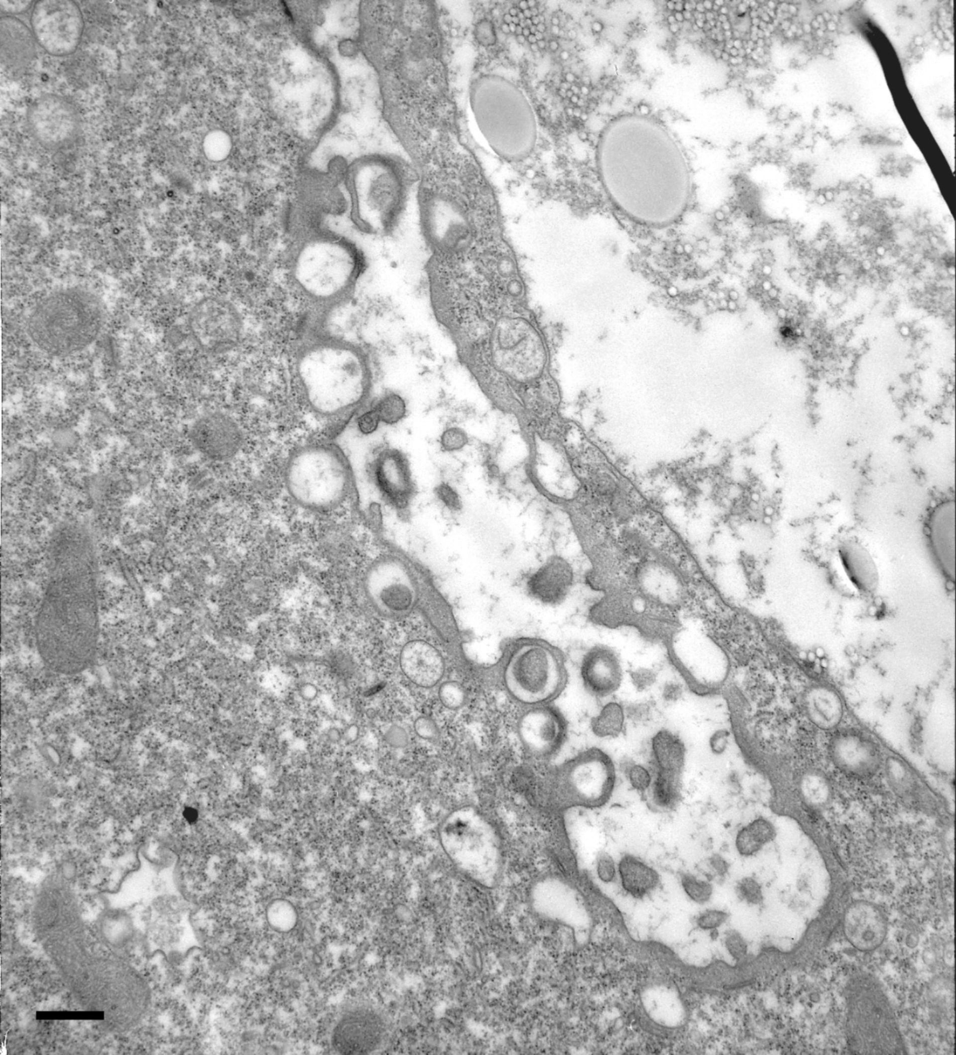
In more extreme cases microfilamentous material builds up into a thick layer surrounding the DV-I membrane as well as the fusion sites between the acidosomes and the DV-I. A similar thick meshwork was not observed between lysosomes and the DV-II. TEM taken on 5/3/79 by R. Allen with Hitachi HU11A operating at 75kV. Neg. 10,000X. Bar = 0.5?m.
Biological Process: Digestive system process
Standard glutaraldehyde fixation followed by osmium tetroxide, dehydrated in alcohol and embedded in an epoxy resin. Microtome sections prepared at approximately 75nm thickness. The negative was printed to paper and the image was scanned to Photoshop. This digitized image is available for qualitative analysis. Additional information available at (http://www5.pbrc.hawaii.edu/allen/).
Author: Richard Allen (University of Hawaii)
Source: The Cell: An Image Library