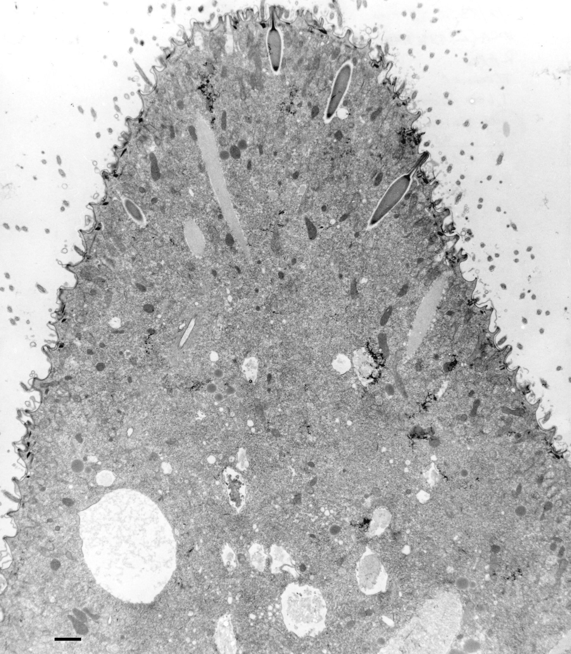
A relatively low magnification view of the anterior end of Paramecium showing the placement and size of trichocysts relative to the cortex and cytoplasmic organelles. Trichocysts docked at the crests of the surface ridges have “carrot” shaped bodies surmounted by a spike that is long enough to penetrate from below the infraciliary lattice all the way to the tip of the ridge. Two trichocysts have been internally discharged into elongated shafts during fixation. cv, contractile vacuole. TEM taken on 12/2/83 by R. Allen with Zeiss 10A operating at 80kV. Neg. 1,600X. Bar = 2?m.
Biological Process: Organelle organization, Trichocyst distribution
Standard glutaraldehyde fixation followed by osmium tetroxide, dehydrated in alcohol and embedded in an epoxy resin. Microtome sections prepared at approximately 75nm thickness. The negative was printed to paper and the image was scanned to Photoshop. This digitized image is available for qualitative analysis. Additional information available at (http://www5.pbrc.hawaii.edu/allen/).
Author: Richard Allen (University of Hawaii)
Source: The Cell: An Image Library