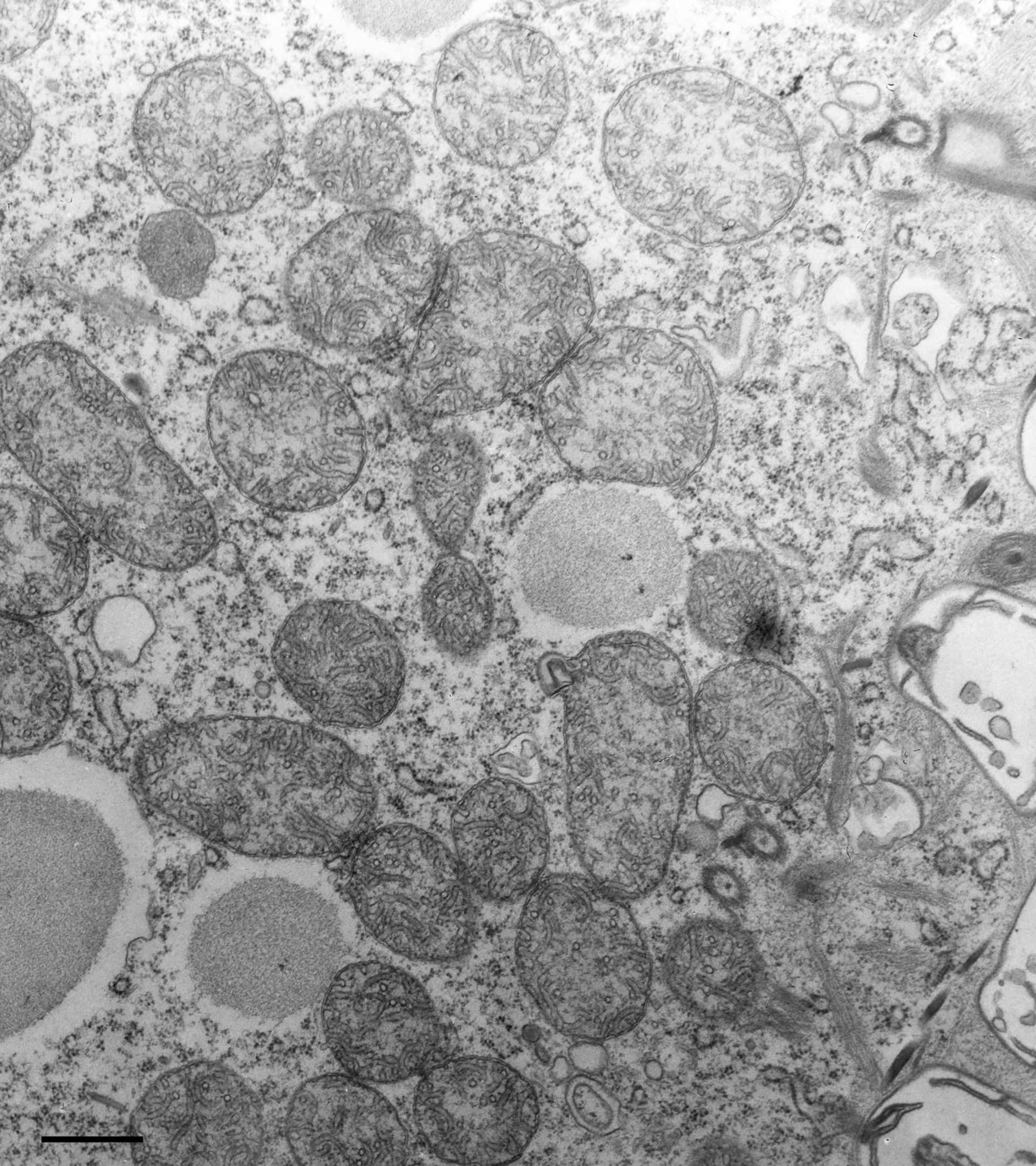
Mitochondria are organelles enclosed by two membranes in which the inner membrane invaginates to form tubular cristae. The mitochondria shown here are typical of cells in late log phase of growth. This cell was fixed 5 days after transferring the cell to fresh culture. Cacodylate-buffered fixative. TEM taken on 5/30/78 by R. Allen with Hitachi HU11A operating at 75kV. Neg. 12,250X. Bar = 0.5?m.
Biological Process: Mitochondrion organization, Cytoplasm organization
Standard glutaraldehyde fixation followed by osmium tetroxide, dehydrated in alcohol and embedded in an epoxy resin. Microtome sections prepared at approximately 75nm thickness. The negative was printed to paper and the image was scanned to Photoshop. This digitized image is available for qualitative analysis. Additional information available at (http://www5.pbrc.hawaii.edu/allen/).
Author: Richard Allen (University of Hawaii)
Source: The Cell: An Image Library