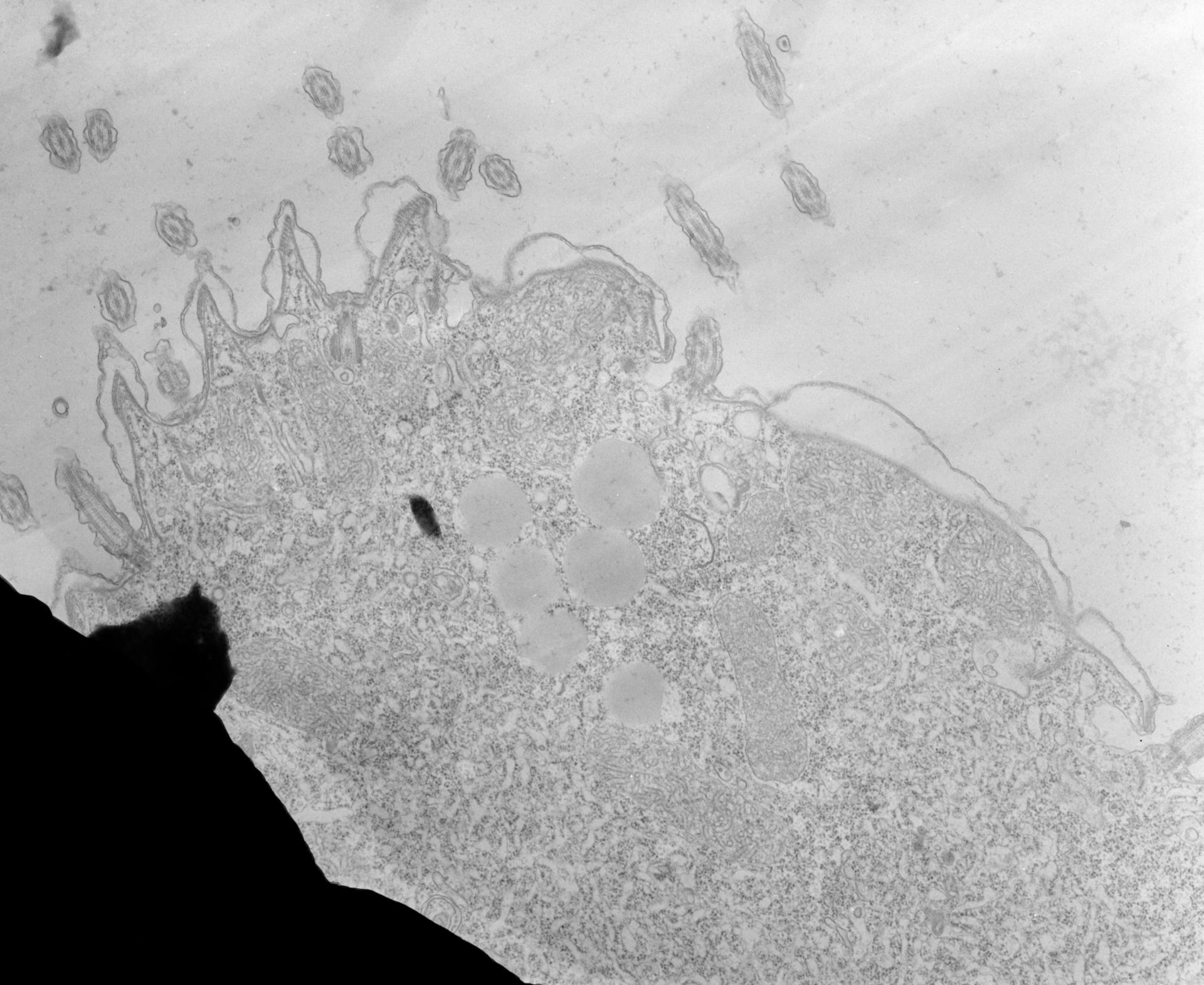
High resolution image showing that one parasomal sac lies anterior to each basal body of the somatic ciliature. A parasomal sac is an indentation of the plasma membrane that penetrates into the cytosol past the alveolus. Most sacs end as a coated pit. However, as seen in this micrograph, they can also give rise to more extensive tubules and vesicles. These vesicles pinch off into the cytosol to become early endosomes in a process called endocytosis where they lose their coat and fuse with a flattened smooth-membraned cisterna that is the early endosome. TEM taken on 7/17/67 by R. Allen with Philips 200 operating at 60kV. Neg. 9,000X. The raw film was scanned with an Epson Perfection V750 Pro. This image is best used for quantitative analysis.
Biological Process: Cortical cytoskeleton organization, Cargo loading into COPII-coated vesicle, Clathrin coat assembly
Standard glutaraldehyde fixation followed by osmium tetroxide, dehydrated in alcohol and embedded in an epoxy resin. Microtome sections prepared at approximately 75nm thickness. Additional information available at (http://www5.pbrc.hawaii.edu/allen/).
Author: Richard Allen (University of Hawaii)
Source: The Cell: An Image Library