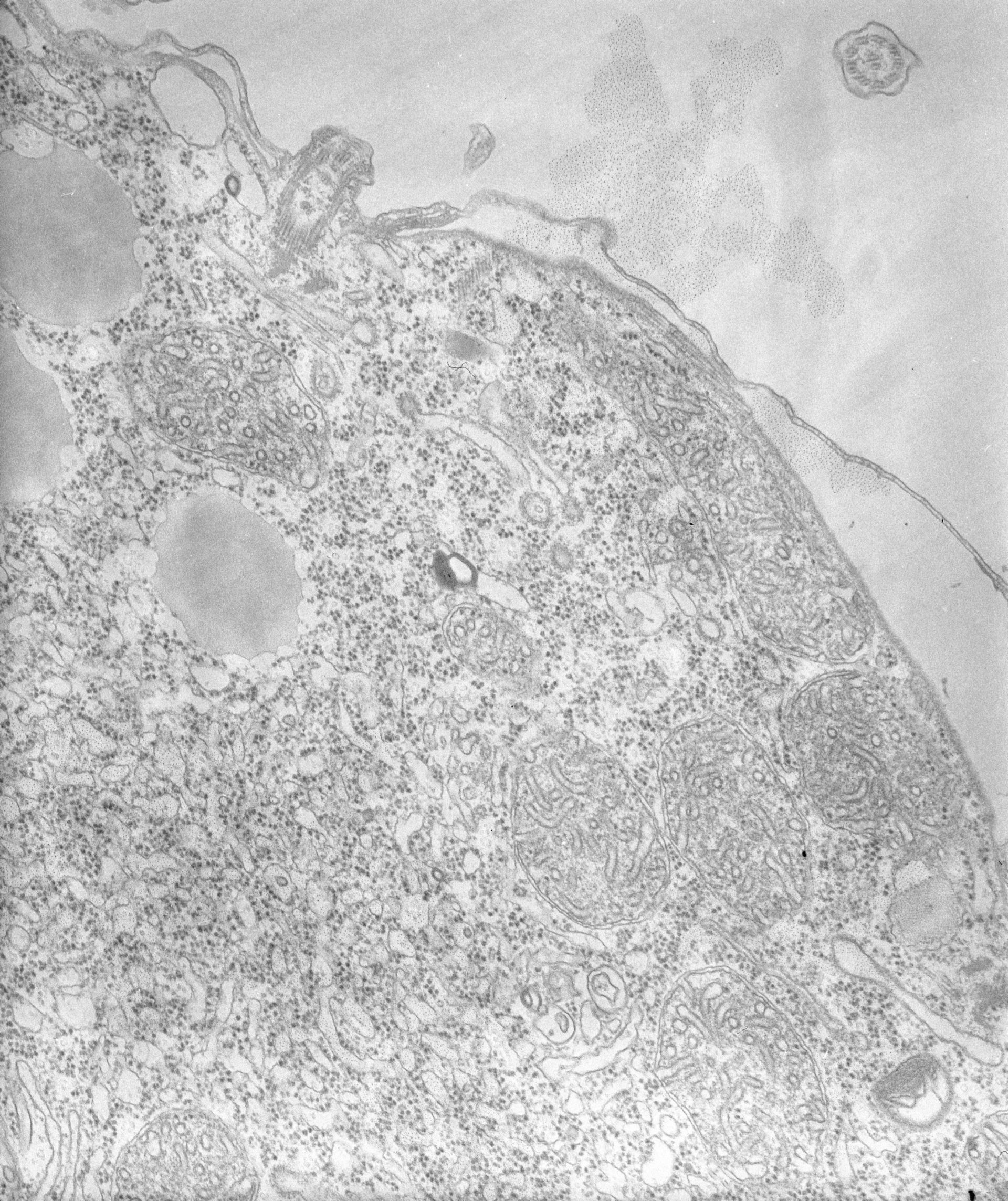
High resolution view of early endosomes with coated evaginations at their margins.TEM taken on 8/15/67 by R. Allen with Philips 200 operating at 60kV. Neg. 19,200X. The raw film was scanned with an Epson Perfection V750 Pro. This image is best used for quantitative analysis.
Standard glutaraldehyde fixation followed by osmium tetroxide, dehydrated in alcohol and embedded in an epoxy resin. Microtome sections prepared at approximately 75nm thickness. Additional information available at (http://www5.pbrc.hawaii.edu/allen/).
Author: Richard Allen (University of Hawaii)
Source: The Cell: An Image Library