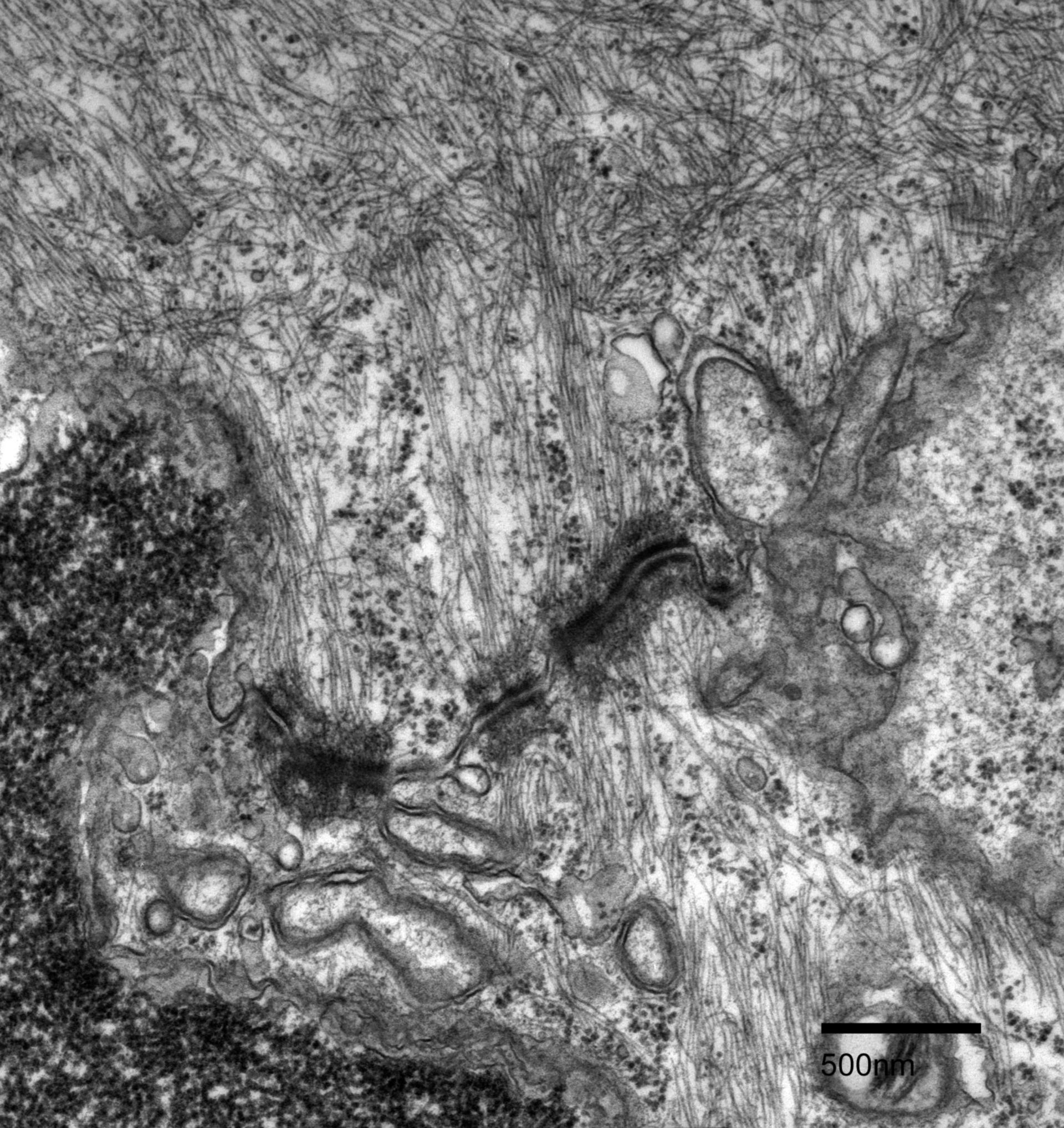
Cell junctions formed between adjacent epidermal cells in Fundulus heteroclitus scales. Desmosomes line the membranes of two epidermal cells. A vast network of intermediate filaments and microfilaments are present throughout the cytoplasm and connect to the desmosomes plates. Microtubules are also visible in the cells.
Biological Process: Cytoskeleton organization, Cell-cell junction organization
Fundulus heteroclitus scales were chemically fixed with 2.5% glutaraldehyde, 2% formaldehyde in 0.1M cacodylate buffer (pH 7.3), then post-fixed in 4% osmium tetroxide and stained en bloc in 1% uranyl acetate. The scales were then dehydrated in a graded series of ethanol and infiltrated with Spurr’s resin. Thin sections of 70 nm were trimmed using a diamond knife and post-stained in uranyl acetate and lead citrate. This micrograph was imaged using a Phillips CM 100 transmission electron microscope at an accelerating voltage of 80 kV.
Author: Marian Rice
Source: The Cell: An Image Library