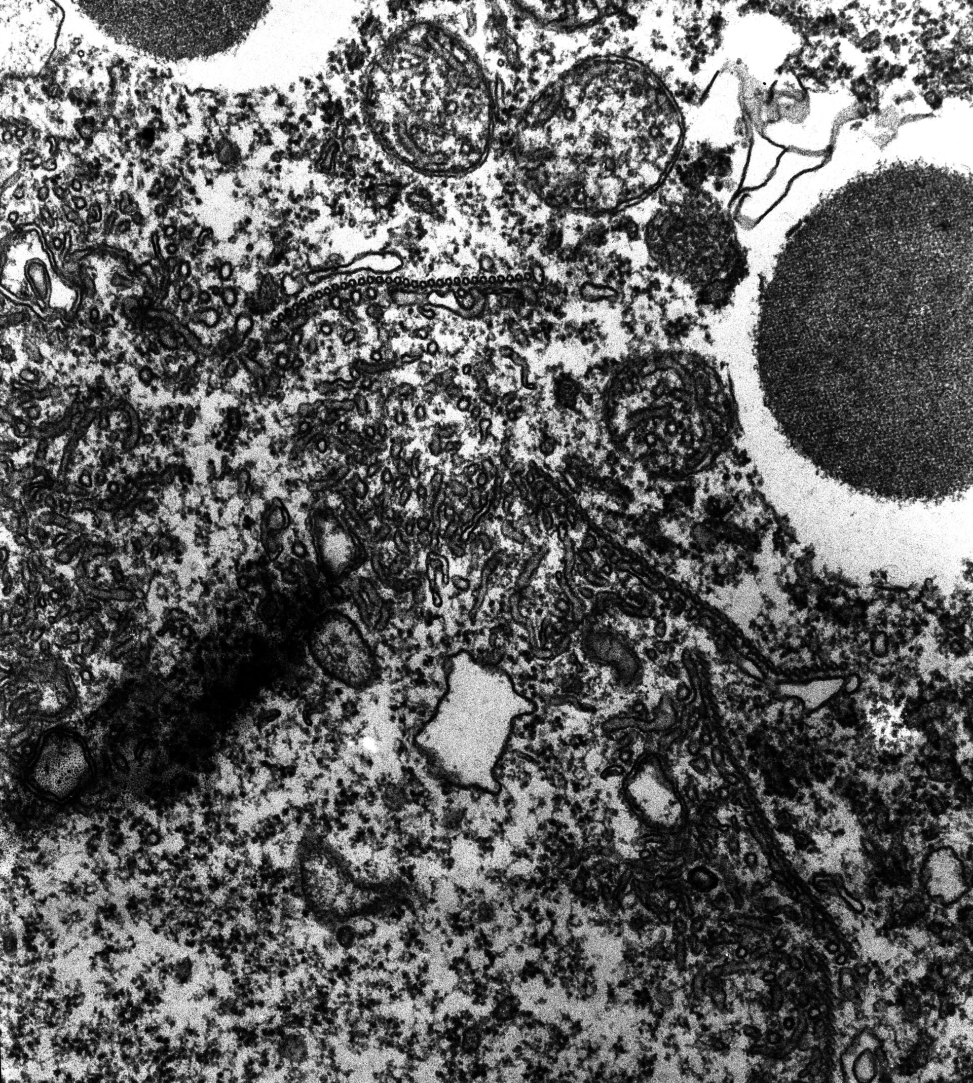
A high resolution view of the tubulated CV membrane attached to ampullae 2, 3, 4 and 5 of the CVC. The tubules lie against the ribbon of microtubules as well as form a large mat lying inward (centrifically) from the ribbons. These tubular CV membranes are part of the smooth spongiome. TEM taken on 2/26/80 by R. Allen with Hitachi HU11A operating at 75kV. Neg. 22,500X.The raw film was scanned with an Epson Perfection V750 Pro. This image is best used for quantitative analysis. Standard glutaraldehyde fixation followed by osmium tetroxide, dehydrated in alcohol and embedded in an epoxy resin. Microtome sections prepared at approximately 75nm thickness. Additional information available at (http://www5.pbrc.hawaii.edu/allen/).
Biological Process: Water transport, Microtubule-based process, Membrane based process
Author: Richard Allen (University of Hawaii)
Source: The Cell: An Image Library