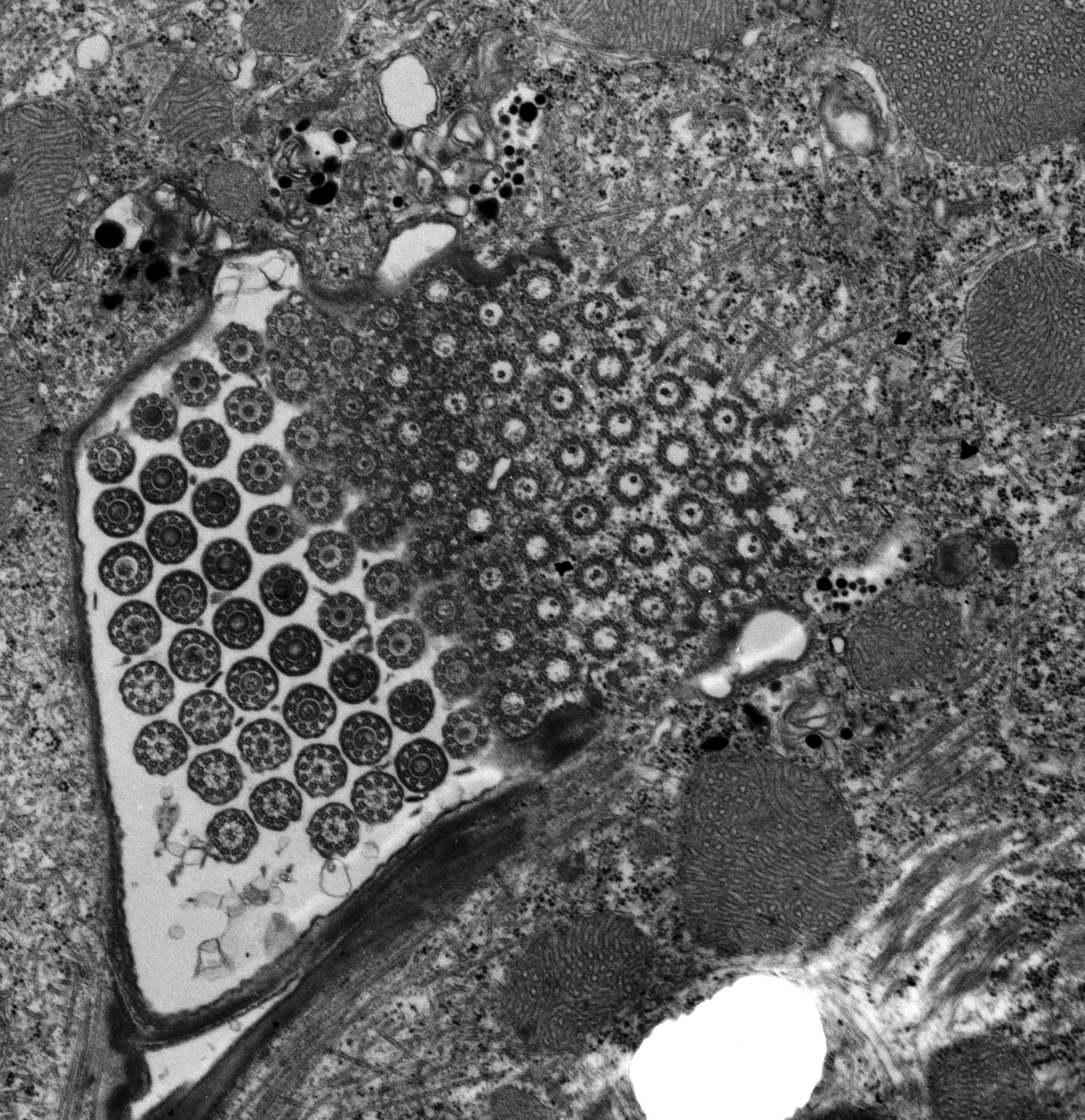
A high resolution image of an individual cirrus of Euplotes with 91 cilia/basal body complexes. The transition from basal body to cilium can be followed from right to left in each row. Small vesicles, many containing electron opaque granules, surround the base of the cirrus. Standard glutaraldehyde fixation followed by osmium tetroxide, dehydrated in alcohol and embedded in an epoxy resin. Microtome sections prepared at approximately 75nm thickness. TEM taken on 7/24/67 by R. Allen with Philips 200 operating at 60kV. Neg. 12,400X. The raw negative was scanned with an Epson Perfection V750 Pro. This image is best used for quantitative analysis. Additional information is available at (http://www5.pbrc.hawaii.edu/allen/).
Biological Process: Ciliary or flagellar motility, Ciliary cell motility, Cytoplasmic microtubule organization
Author: Richard Allen
Source: The Cell: An Image Library