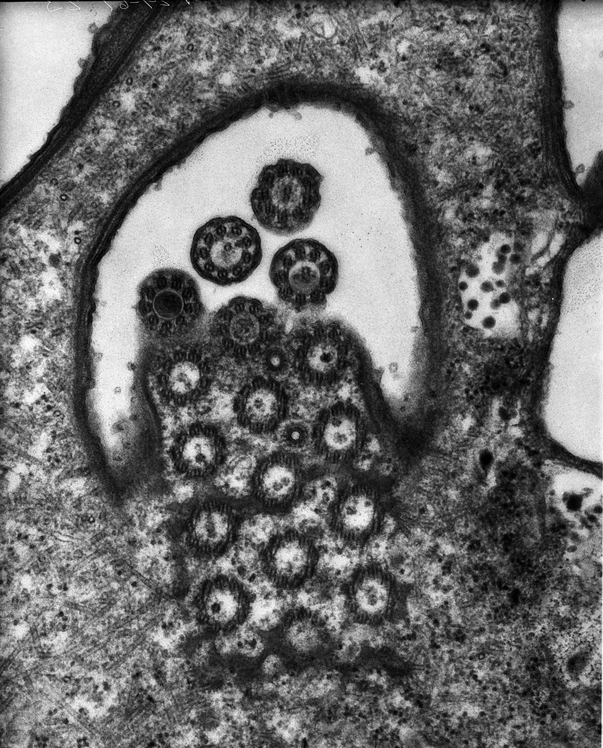
A high resolution image of the transition from basal bodies to cilia within a portion of one of the oral membranelles of Euplotes. Parasomal sacs occur between the rows of basal bodies although these endocytic ports are not as evident in Euplotes as in other ciliates. Standard glutaraldehyde fixation followed by osmium tetroxide, dehydrated in alcohol and embedded in an epoxy resin. Microtome sections prepared at approximately 75nm thickness. TEM taken on 7/29/67 by R. Allen with Philips 200. Neg. 28,000X. The raw negative was scanned with an Epson Perfection V750 Pro. This image is best used for quantitative analysis. Additional information is available at (http://www5.pbrc.hawaii.edu/allen/).
Biological Process: Ciliary or flagellar motility, Ciliary cell motility, Membranelle orgnization
Author: Richard Allen
Source: The Cell: An Image Library