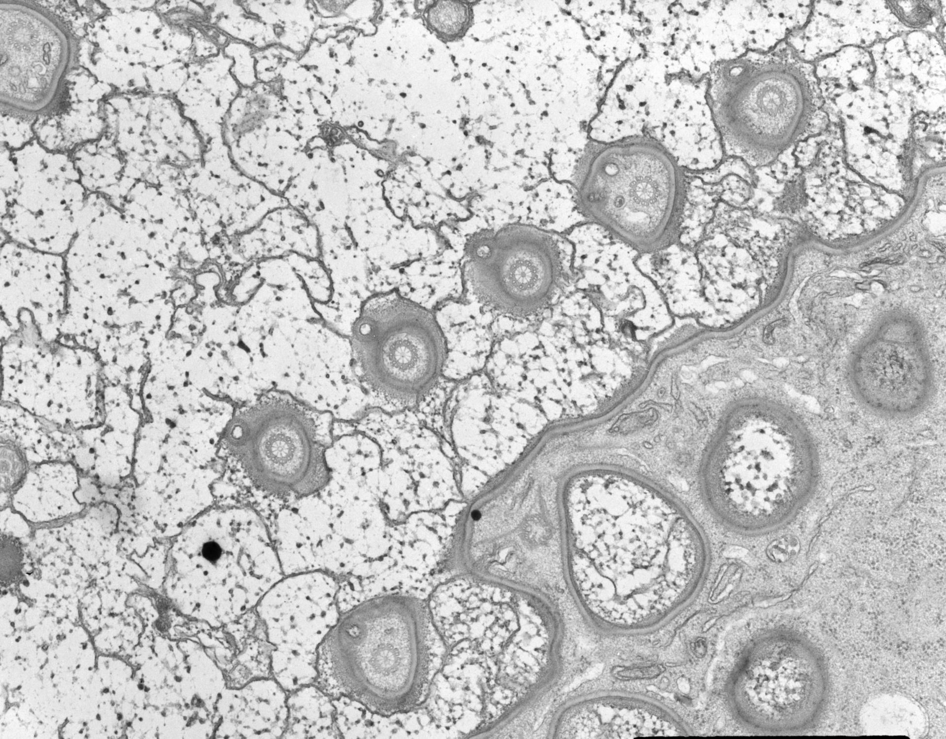
A high resolution view of cross-sectioned rows of basal bodies at the cortex of Nassula. Only a few basal bodies may have attached cilia as seen by the few cilia present in the upper right row. Basal bodies extend to the tips of the cytoplasmic mounds and each mound has a parasomal sac associated to one side. Unlike many other ciliates, the cartwheel occupies most of the basal body lumen. TEM taken on 4/1/69 by R. Allen with Philips 300 operating at 60kV. Neg. 14,800X. The raw film was scanned with an Epson Perfection V750 Pro. This image is available for quantitative analysis. Standard glutaraldehyde fixation followed by osmium tetroxide, dehydrated in alcohol and embedded in an epoxy resin. Microtome sections prepared at approximately 75nm. Additional information available at (http://www5.pbrc.hawaii.edu/allen/).
Biological Process: Cortical cytoskeleton organization, Microtubule cytoskeleton organization, Ciliary or flagellar motility
Author: Richard Allen
Source: The Cell: An Image Library