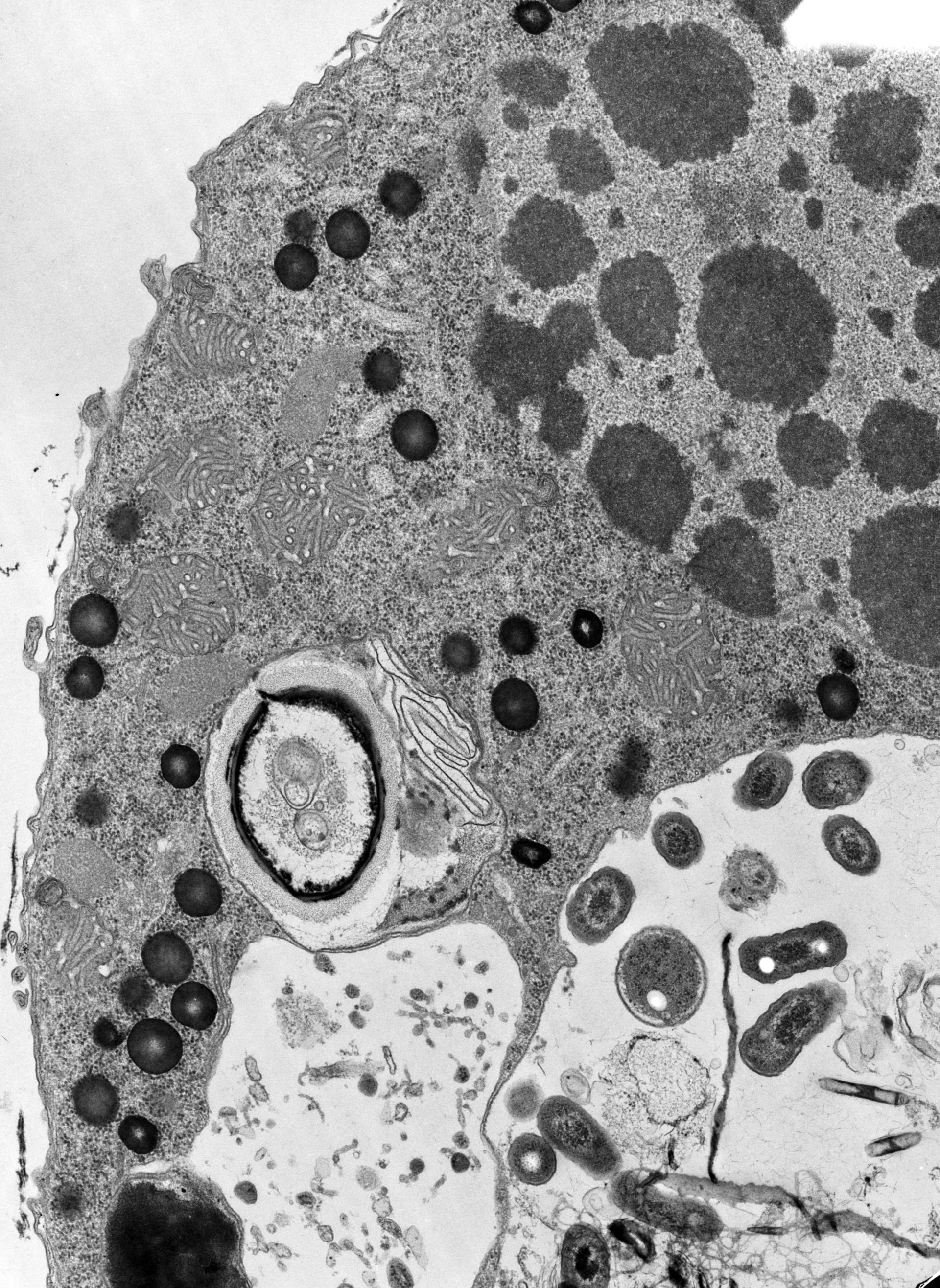
High resolution image of Halteria fixed in cacodylate-buffered fixative and stained. The round, dark bodies, possibly pigment granules, which were transparent in other preparations are electron-opaque following this protocol. The alveoli are flattened and inconspicuous. There is little evidence of an epiplasm. TEM taken on 11/8/68 by R. Allen with Philips 300 operating at 60kV. Neg. 12,500X. The raw film was scanned with a Nikon Coolscan 9000ED. This image is suitable for quantitative analysis. Standard glutaraldehyde fixation followed by osmium tetroxide, dehydrated in alcohol and embedded in an epoxy resin. Microtome sections prepared at approximately 75nm thickness. Additional information available at (http://www5.pbrc.hawaii.edu/allen/).
Biological Process: Macronucleus organization, Cellular pigmentation, Digestion
Author: Richard Allen
Source: The Cell: An Image Library