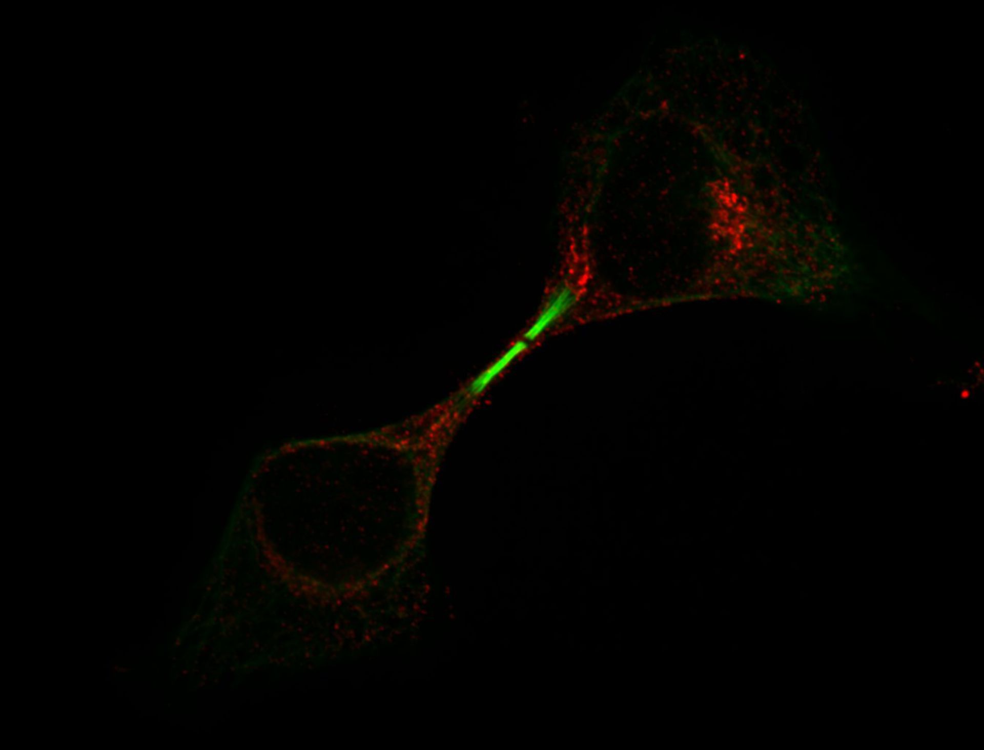
Distribution of Golgi Apparatus during cell division. Hela cells fixed with MeOH on ice for 3 min, and stained with a primary antibody against the Golgi and FITC conjugated DM1a antibody (green) against microtubules. Secondary antibody was Alexa 594 for the Golgi (red). A single section was collected with a spinning disk microscope on a Nikon TE-2000 with a 1.3 NA 100X objective. Images were collected with an Orca ER CCD camera. Lasers and filters: Innova 70C Spectrum 3 watt Laser, 488 Laser/ATOF 525/50, and 568 Laser/ATOF 605/52.
Biological Process: Cell division
Author: Hu, Chi-Kuo
Source: The Cell: An Image Library