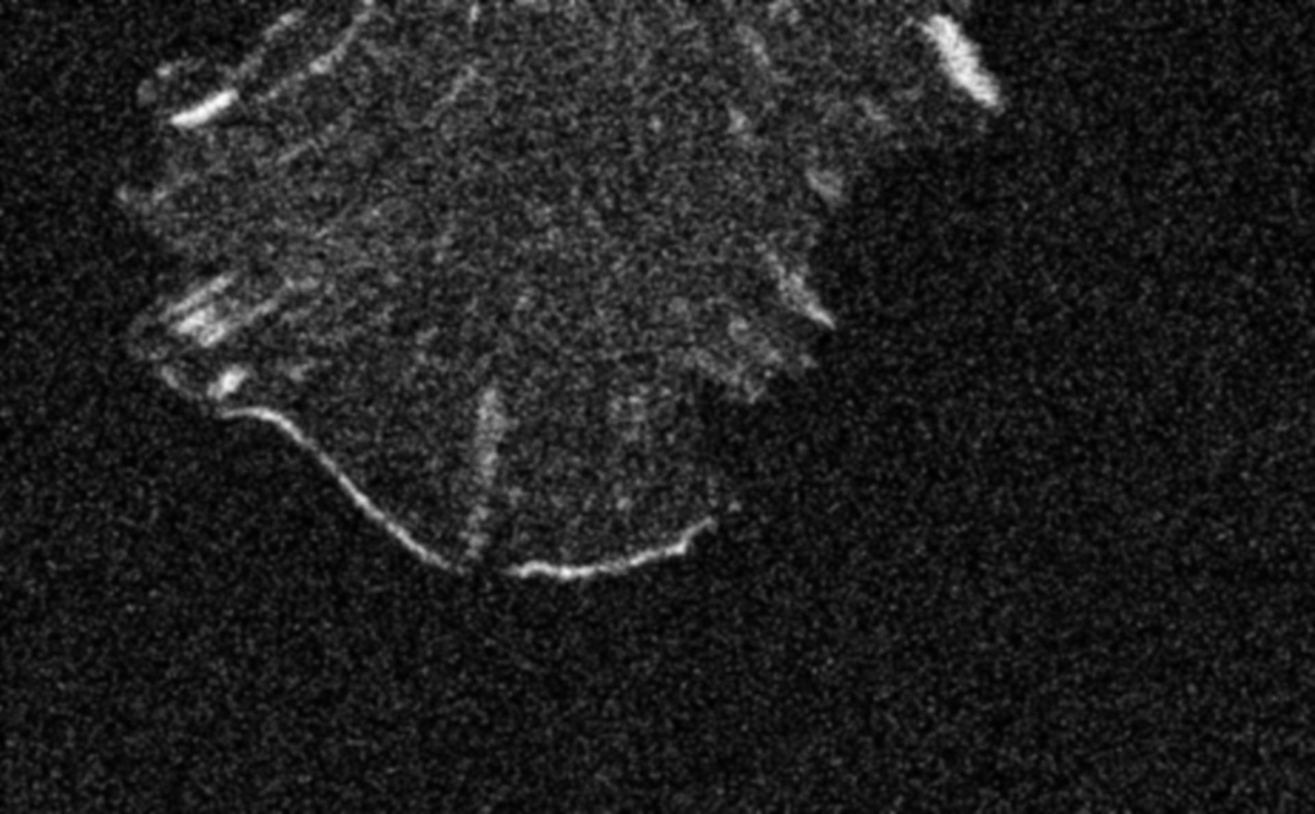
NIH 3T3 cell transfected with EGFP-VASP. VASP is localized to the focal adhesions and is also present along the protruding leading edge. VASP only hightlights the portions of the periphery that are advancing forward, giving VASP a non-uniform distribution along the leading edge. Time series taken as a single confocal slice using a Zeiss LSM 510 confocal with a 100X 1.3 NA objective, 488 nm excitation and a 505-530 BP emission filter.
Biological Process: Regulation of actin cytoskeleton organization
Author: Catherine Galbraith,
Source: The Cell: An Image Library