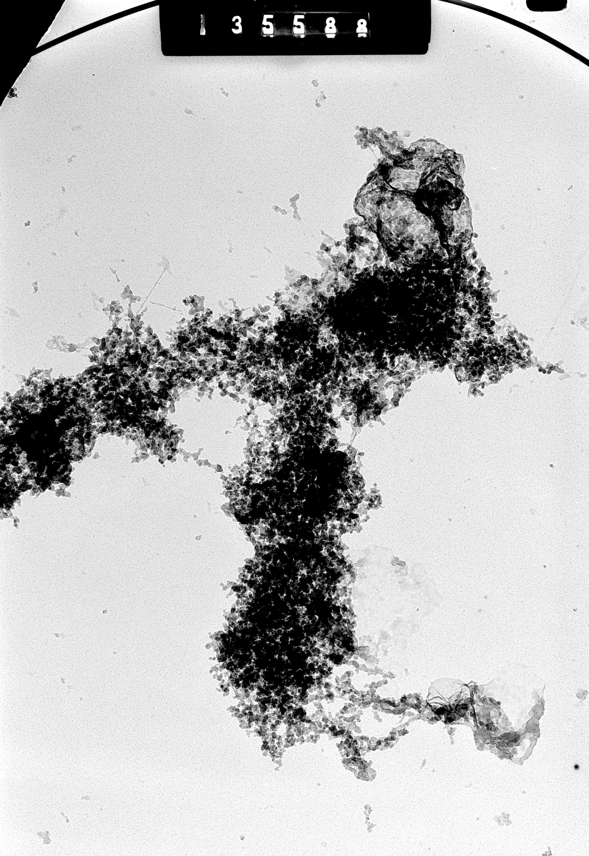
High Voltage (1MeV) EM image of metaphase arrested CHO cell isolated using a non-aqueous technique, spread on a buffer surface and transferred to an EM grid. A single chromatid pair is seen in the center of the image. This micrograph was captured at a specimen tilt of 50 degrees. The accompanying image has a tilt of 60 degrees, creating a stereo pair that provides an oblique stereo view of the chromatids.
Biological Process: Mitosis, Mitotic metaphase, Chromosome organization
Cells were metaphase arrested with colcemid, then exposed to a low ionic strength medium (0.07M phosphate buffer). Chromosomes were prepared by a non-aqueous technique, spread on the surface of a low ionic strength buffer, transferred to an EM grid for examination in the HVEM. See also: H. Ris 1981 Stereoscopic electron microscopy of chromosomes. Meth Cell Biol 22:77-96 H. Ris 1978 Preparation of chromatin and chromosomes for electron microscopy. Meth Cell Biol 18:220-246.
Author: Hans Ris
Source: The Cell: An Image Library