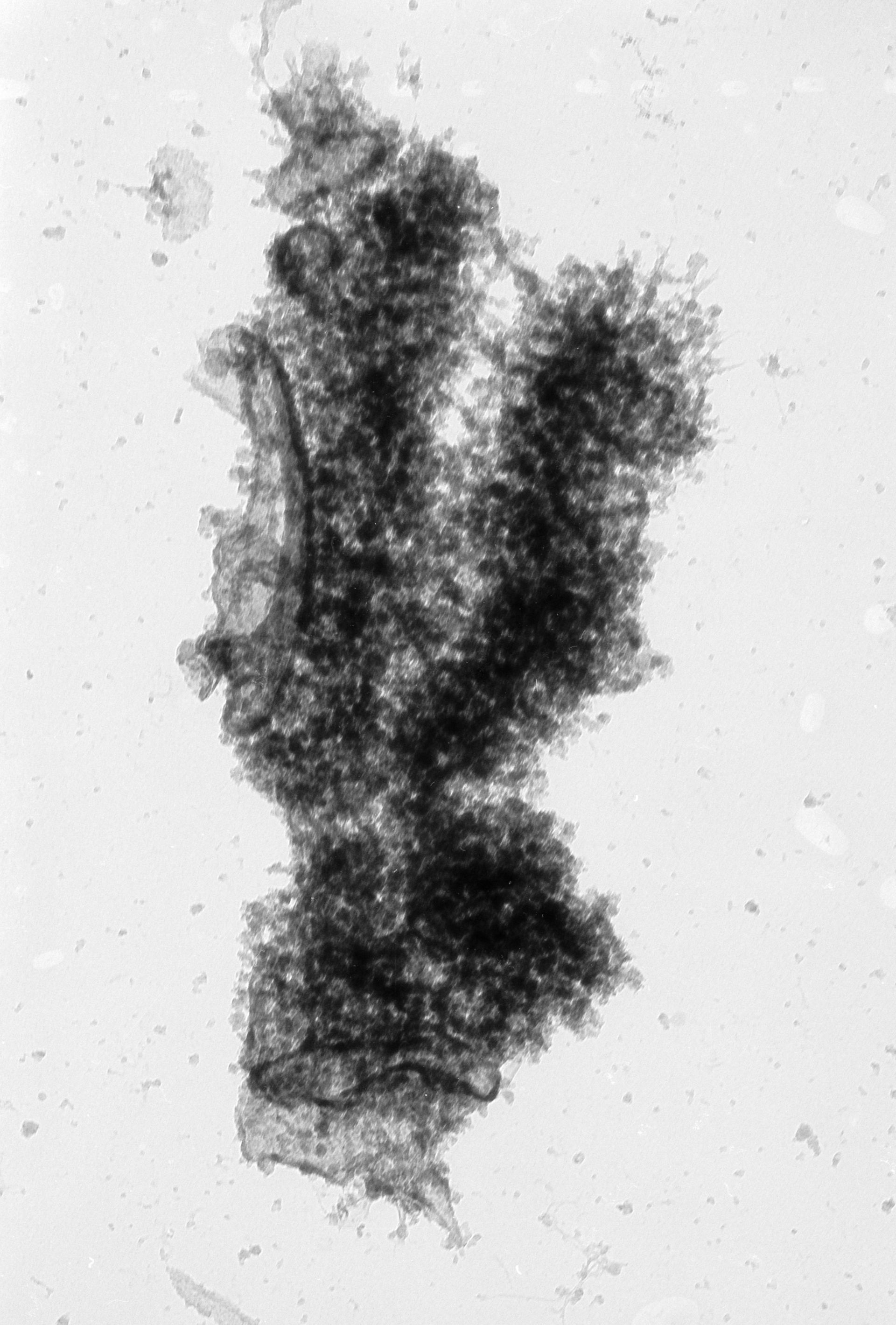
High voltage (1MeV) transmission electron microscopy image of an isolated metaphase chromatid pair from a Chinese hamster ovary cell, showing the fiber-like structure and two large pieces of attached membrane. The image was taken with a specimen tilt of 55 degrees. Grouped with it is an image with a tilt of 48 degrees, providing a pair that affords an oblique stereo view of the chromosome.
Biological Process: Chromosome organization, Mitosis, Mitotic metaphase
Cells were treated with colchicine for 24 hr, and metaphase chromosomes isolated using the non-aqueous hexylene glycol protocol developed by Stubblefield and Wray, placed on a grid, critical point dried, and stained with uranyl acetate. See also: H. Ris 1981 Steroscopic electron microscopy of chromosomes. Meth Cell Biol 22:77-96 H. Ris 1978 Preparation of chromatin and chromosomes for electron microscopy. Meth Cell Biol 18:220-246.
Author: Hans Ris
Source: The Cell: An Image Library