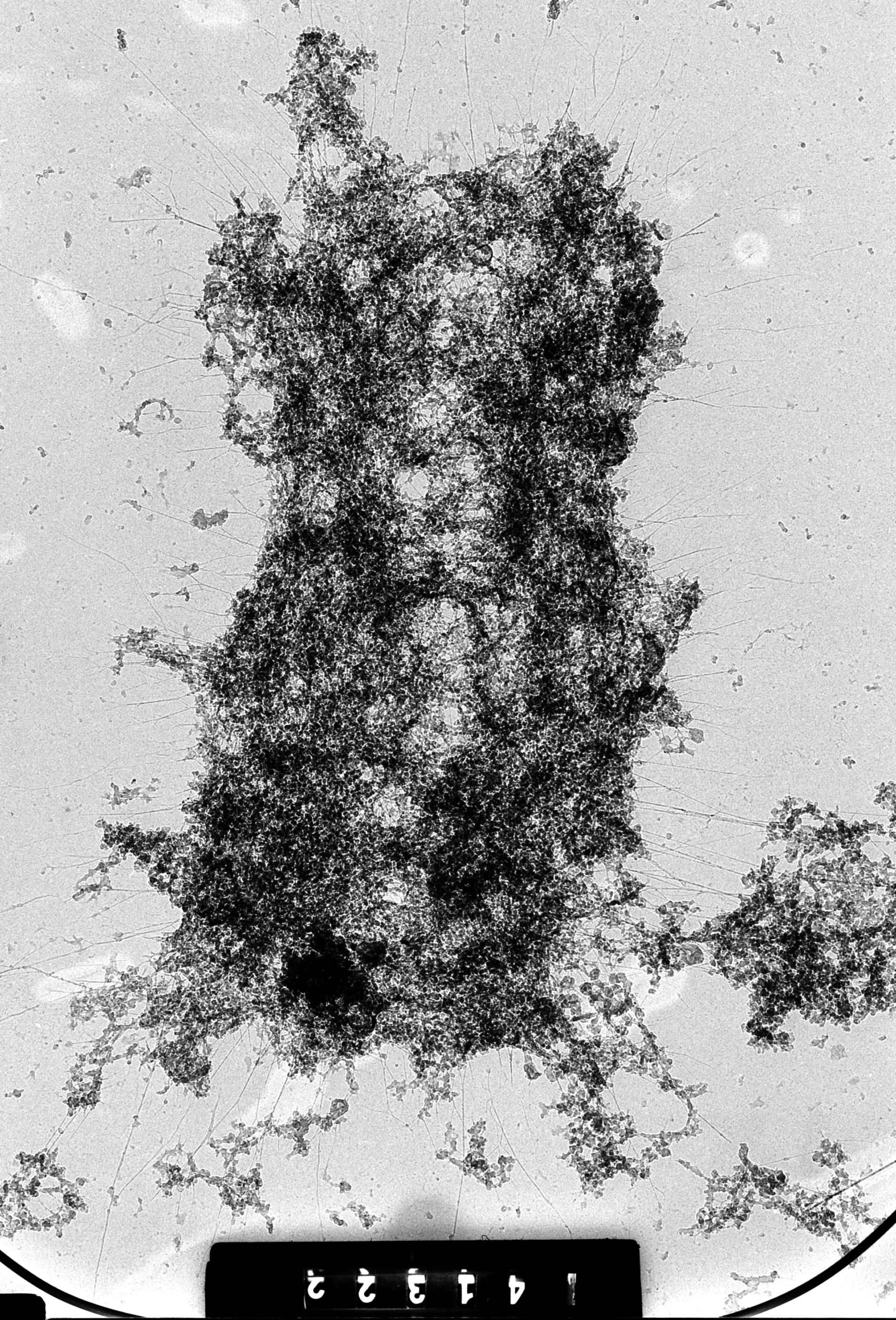
Whole human female metaphase chromosome from metaphase arrested cell swollen in hypotonic medium, and treated to remove histones. The residual 'scaffold' was imaged with high voltage EM at 1 MeV. This image was taken with a specimen tilt of 46 degrees. Grouped with it is an image of the same area at 54 degrees tilt, providing an oblique stereo view of the chromosome.
Biological Process: Nuclear division
HeLa cells were metaphase-arrested by a 3hr treatment with colcemid, the cells mechanically broken by passage through a 22-gauge needle and exposed to hypotonic 75 mM KCl, treated to remove histones, spread on a water surface, fixed with formaldehyde, picked up on formvar coated grid, critical point dried, and imaged with the Wisconsin HVEM at 1 MeV. See Ris (1978) Preparation of chromatin and chromosomes for electron microscopy. Meth Cell Biol 18:229-246; Ris (1981) Stereoscopic microscopy of chromosomes. Meth Cell Biol 22:77-96.
Author: Hans Ris
Source: The Cell: An Image Library