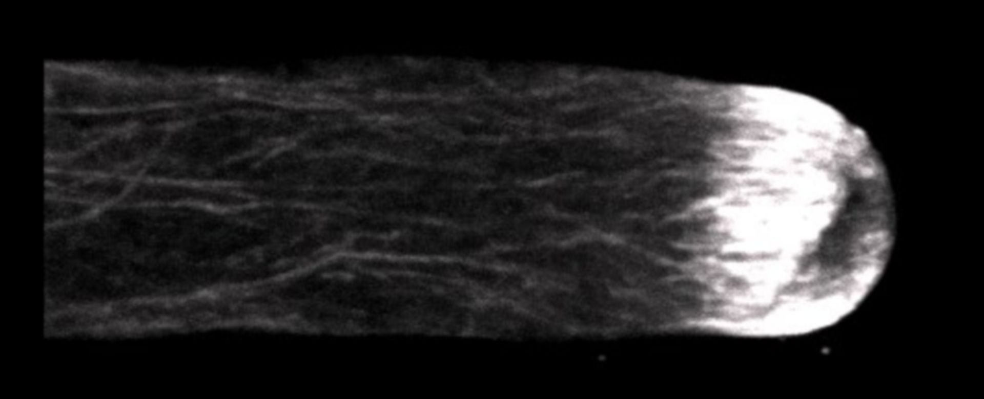
Freeze substituted Lilium pollen tube immunolabeled for filamentous actin and imaged using laser scanning confocal microscopy. Shown is a projection of z-slices revealing the dense sub-apical actin fringe. Other images in the group reveal the distributions of other cytoskeletal elements and membranous components.
Biological Process: Pollen tube growth
Germinated lily pollen on agar loops was plunge frozen, freeze substituted, labeled with an anti-actin mouse monoclonal antibody followed by Cy-3 goat anti-mouse and imaged with a Zeiss 510 meta confocal microscope using a 63x 1.4 NA objective lens. The image shown is a projection of z-slices. See Lovy-Wheeler et al. 2005, Planta 221:95-104.
Author: Peter K Hepler
Source: The Cell: An Image Library