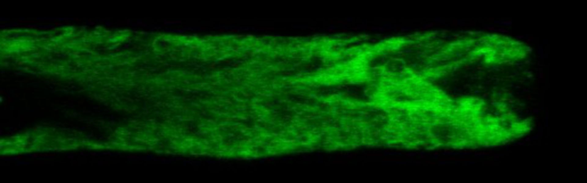
Living Lilium pollen tube labeled with mGFP5-HDEL to mark endoplasmic reticulum (green) imaged using laser scanning focal microscopy. Shown is a central 1.0 micron thick x-y slice. The growing tip is devoid of both organelles. Other images in the group reveal the distributions of additional cytoplasmic components.
Biological Process: Pollen tube growth
Living lily pollen tubes were labeled by bombardment with tungsten bullets coated with plasmid DNA containing the mGFP5-HDEL gene and, after expression, imaged with a Zeiss 510 meta confocal microscope using a 63x 1.4 NA objective lens. The signal from a central x-y plane through the tube is shown. See Lovy-Wheeler et al. 2007 Cell Motil Cytoskel 64:217-321.
Author: Peter K Hepler
Source: The Cell: An Image Library