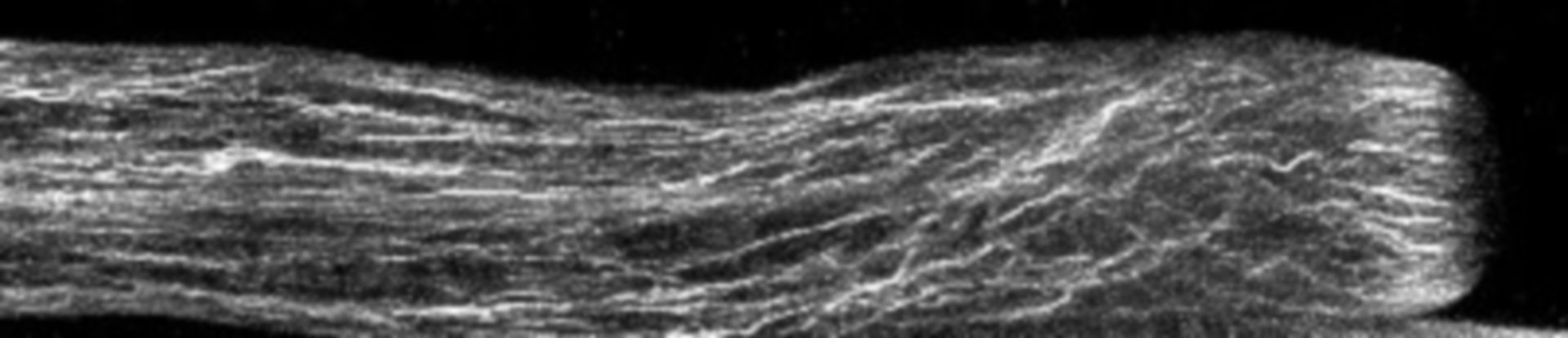
Fixed Lilium pollen tubes were treated with fluorescent phalloidin to label filamentous actin, and confocal images obtained. A strongly labeled actin fringe near the growing tip is present. Other images in the group reveal the distributions of additional cytoskeletal elements and membranous components.
Biological Process: Pollen tube growth
Germinated lily pollen was fixed with paraformaldehyde and glutaraldehye, treated with Alexa-543 phalloidin to label actin, and imaged with a Zeiss 510 meta confocal microscope using a 63x 1.4 NA objective lens. Image is a projection of several confocal x-y slices. See Lovy-Wheeler et al. 2005, Planta 221:95-104.
Author: Peter K Hepler
Source: The Cell: An Image Library