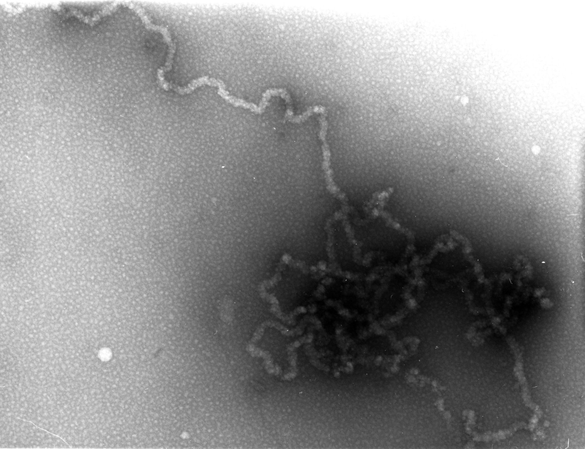
Newt (Notopthalmus viridenscens erythrocytes were isolated in 0.1M KCl, spread on the surface of Na-citrate, picked up on formvar-carbon films, fixed with paraformaldehyde and negatively stained with 2% uranyl acetate. Images were recorded at 40KX and accelerating voltage of 1MeV at the University of Wisconsin HVEM facility.This image was taken with a specimen tilt of 60 degrees. A grouped image of the same field at a 40 degree tilt provides an oblique stereo view of ~30 nm chromatin fibers emanating from the dispersed nucleus.
Biological Process: DNA packaging
Author: Hans Ris
Source: The Cell: An Image Library