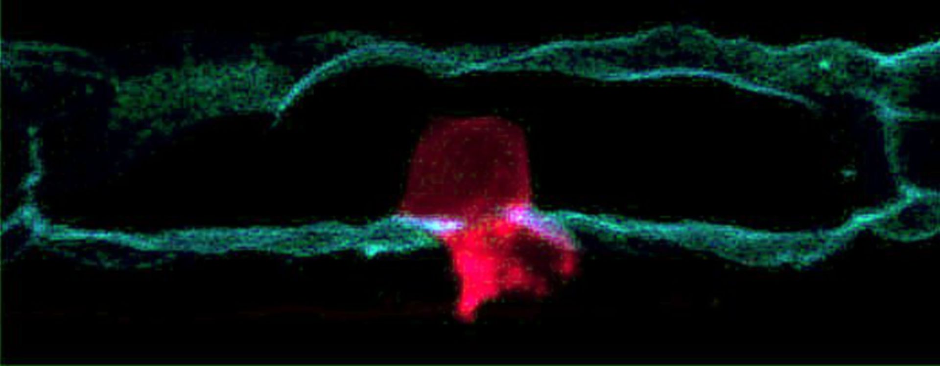
The image shows an anchor cell (red) in the developing female reproductive system of C. elegans pushing through the basement membrane (green) that surrounds it. This type of invasion is also often seen during cancer metastasis.
Biological Process: Cell migration
Anchor cells were labeled with mCherry and laminin with GFP. Images were acquired using a Zeiss AxioImager A1 microscope with a 100X Plan- APOCHROMAT objective and a Zeiss AxioCam MRm CCD camera, controlled by Zeiss Axiovision software (Zeiss Microimaging Inc., Thornwood, NJ). See JW Zeill et al 2009. Nat Cell Biol. 11:183–189.
Author: Eliot Hagedorn
Source: The Cell: An Image Library