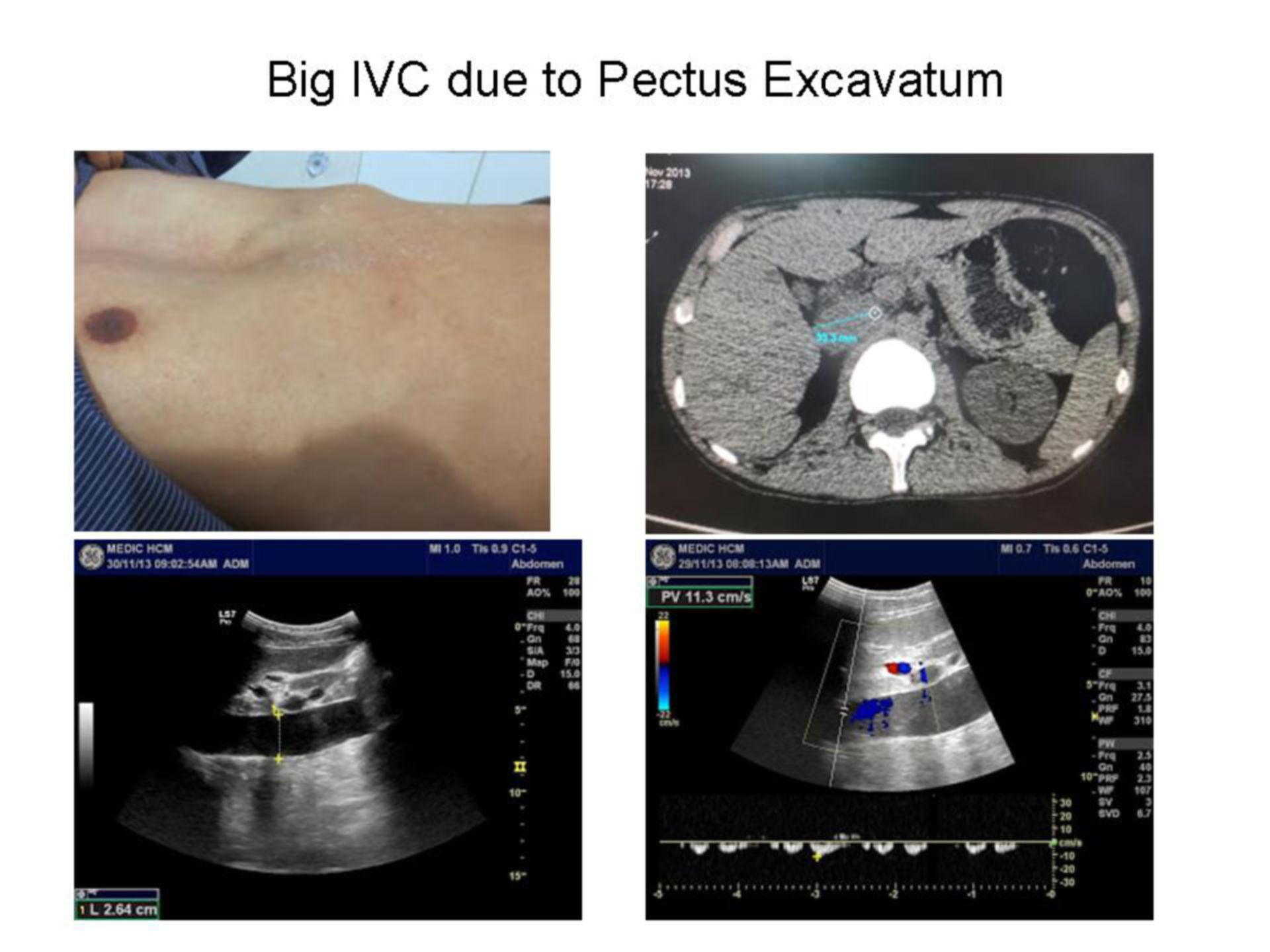
Man 33 yo, pectus excavatum status, onset of epigastric pain with radiating backward todorsal spine. Ultrasound of abdomen detected a big inferior vena cava (IVC) with size of 2.4-2.7cm, echogenic blood flow. Spectrum Doppler of IVC showed triphasic pattern with V= 11.2cm/s, TEE 3D cardiac report was normal. MSCT of abdomen showed that IVC was dilated in going to heart, diameter of 2.9-3.3 cm. Suggested IVC dilated due to PE (pectus excavatum).