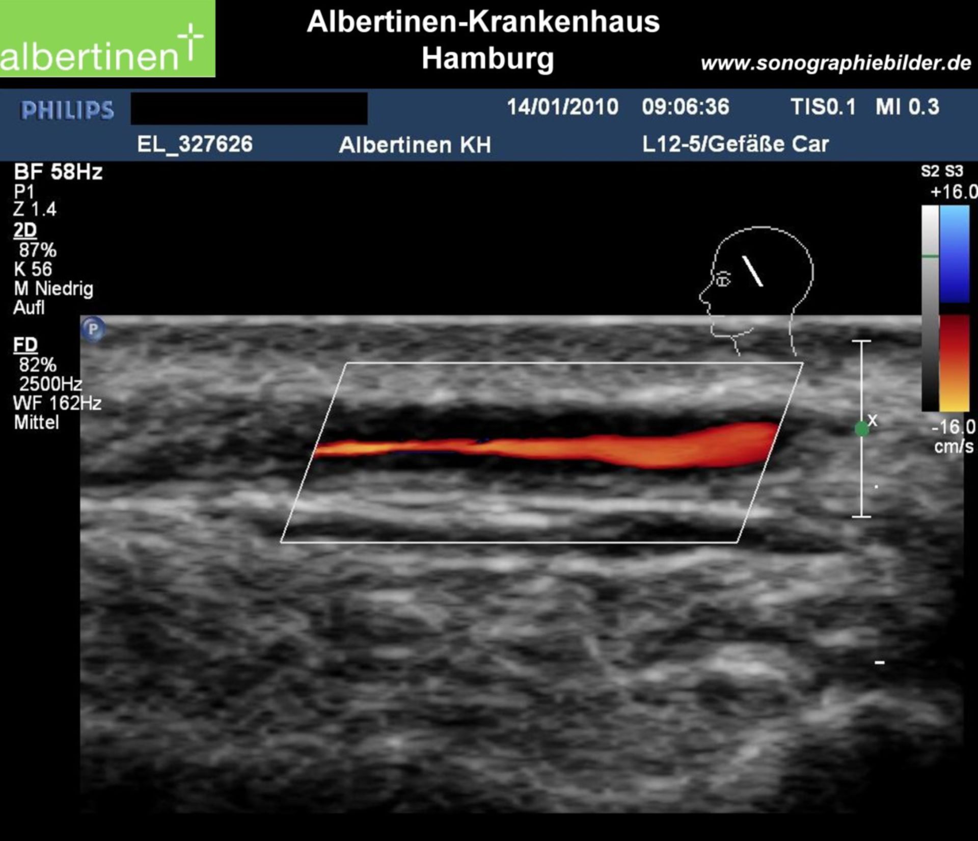
Sonographic image in the longitudinal view of a temporal artery. The hypoechogenic areas (inflammation) are clearly visible. Thickened artery wall, still existing blood flow (duplex sonography).