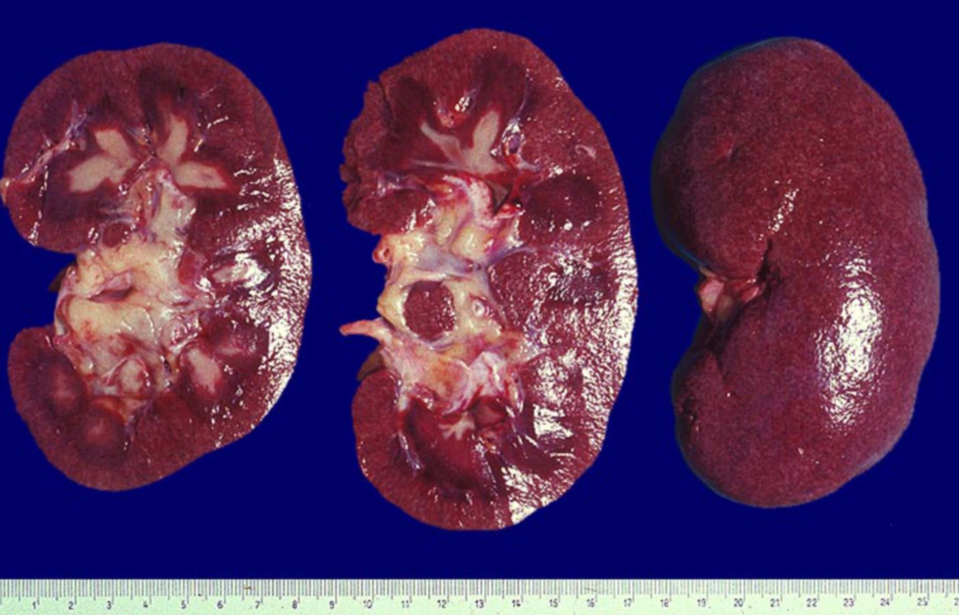
Diagnosis: Lupus erythematodus: papillary necrosis
Description: Enlargement of kidneys. The kidneys surface is slightly spotted. Multiple papillary necrosis and hyperaemia in the border region of the cortex and medulla. Papillary necrosis is a sign of an additional vasculitis.
Commentary: 70% of kidney biopsies of SLE patients are positive for lightmicroscopical changes and in over 90% for electromicroscopically detectable defects. All morphological glomerulonephritis types can occur. Mixed types are frequent. In repeated biopsy probes transient forms from one to another type can be seen.
WHO classification:
(Source: ©PathoPic)