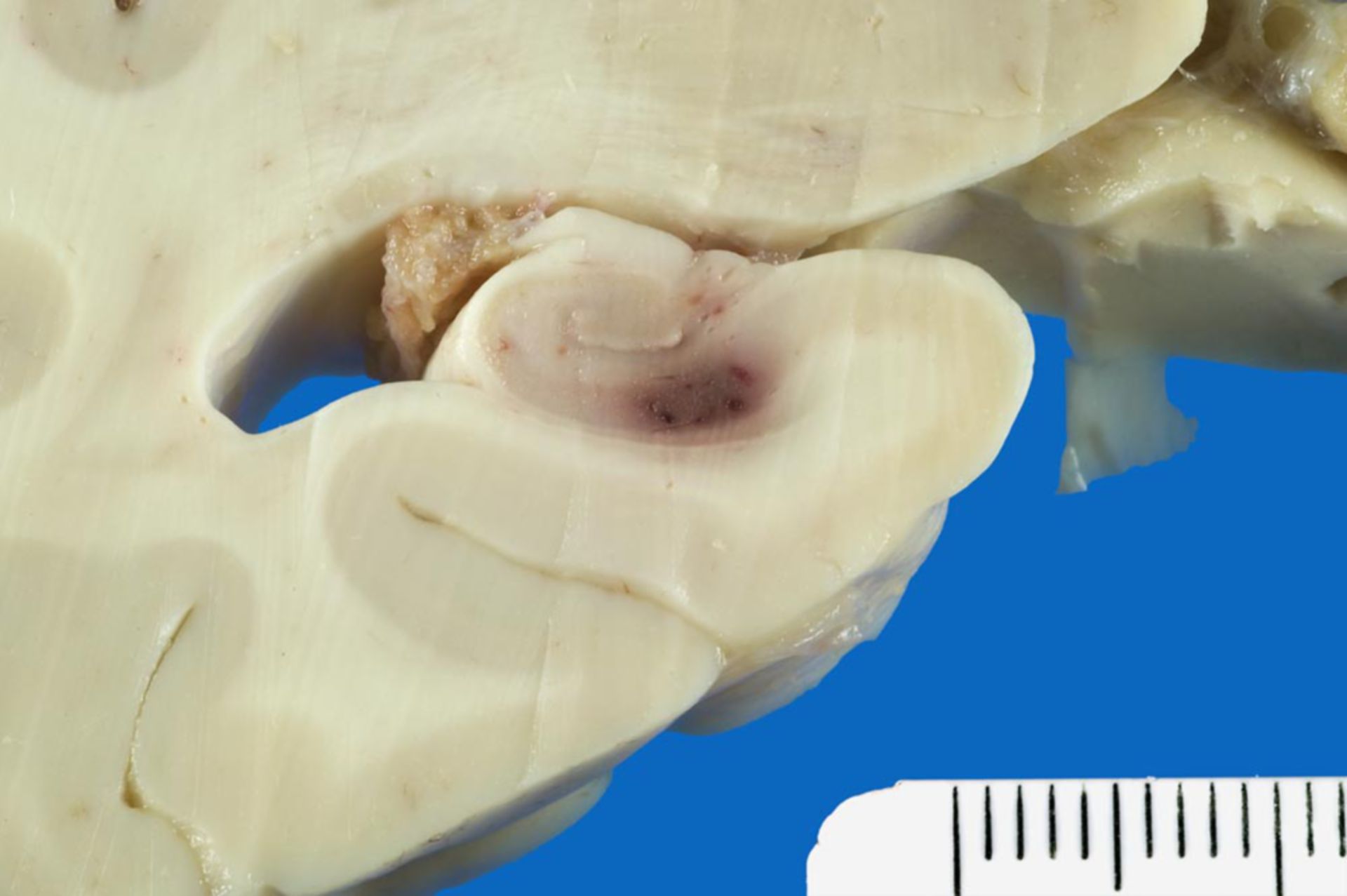
Diagnosis: Petechial bleeding into the hippocampus in HHV-6 infection
Description: Fresh petechial bleeding in the hippocampus.
Additional findings: During the neuropathological autopsy of the brain several macroscipic small, fresh hemorrhages were found in the striatum and also in the thalamus, amygdala and hippocampus.
The microscopic workup of specimen from these regions confirmed the macroscopic impression and additonally showed severe hippocampal sclerosis on both sides. There was subtotal loss of nerve cells in the hilus region and the CA3-region, moderate neurolnal loss in the CA2-region and an almost total loss of nerve cells in the Sommer's secotor (CA1-region). Remarkable was an unusually high reactive astrogiosis in all of the hippocampus region and in the amygdala.
Clinically: 29-year old male patient, acute lymphoblastic leukemia (ALL) known for 3 years. Allogene stem cell transplantation 9 weeks before death.
Commentary: These unusual macroscopic and microscopic findings are compatible with a clinically suspected HHV-6 reactivation after a hematopoetic stem cell transplantation. The occurence of fatal HHV-6 associated encephalits after stem cell transplantation has been described in the literature (see Drobyski WR et al., NEJM, 1994) and there are also reports of hippocampal sclerosis in HHV-6 encephalitis (in status post stem cell transplantation; Wainwright MS et al., Ann Neurol, 2001).
The ocurrance of petechial bleeing into CNS-tissue can be interpreted as a thrombotic microangiopathy and has also been described in literature connected to HHV-6 encephalitis after stem cell transplantation (Belford A et al., Am J Hematol, 2004).
Finally the here present severe astrogliosis in the hippocampus and amygdala can be traced to a recently described tropism of HHV-6 for hippocampal astrocytes (see Fotheringham J et al., J Infect Dis, 2007).
In summary, because of the clinical progression and the neuropathological autopsy and microscipic findings, it can be assumed with high certainty that this is a case of HHV-6 associated hippocampal sclerosis with accompanying also HHV-6 associated bleeding caused by thrombotic microangiopathy, even if PCR-virus-detection cannot be proof because of about 90% seropositivity.
Source: ©PathoPic