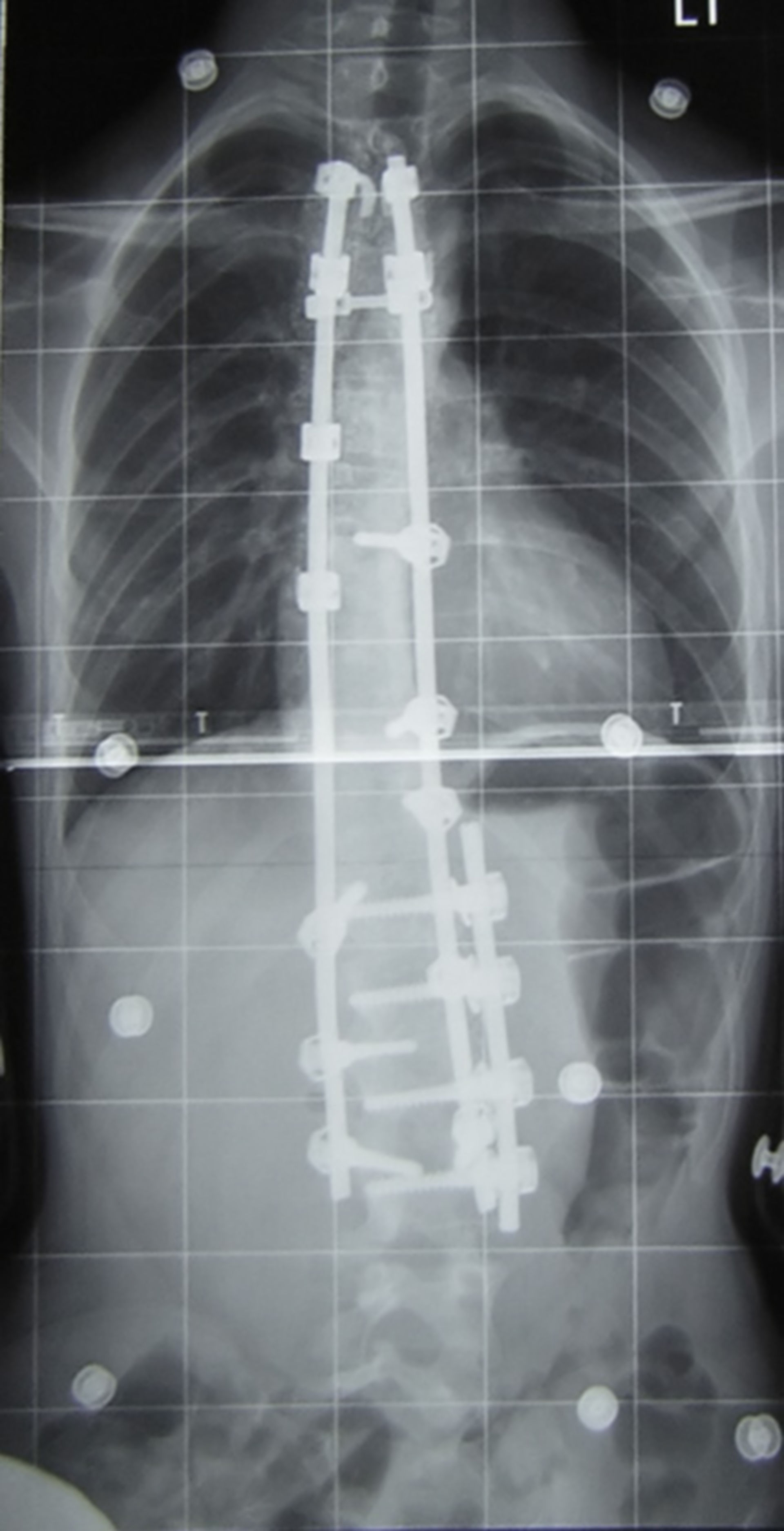
This is an anterior-posterior X-ray of a case of adolescent idiopathic scoliosis post-fusion - specifically, my spine. There was originally a thoracic curve of 30° and a lumbar curve of 53° (Cobb angle - see scoliosis) and these curves have been reduced to less than 15° each.
This was taken at the Royal National Orthopaedic Hospital. The largest curve (53°) is of a magnitude typically near the lower surgery boundary, although many factors decide whether surgery is necessary on a scoliosis case.
This x-ray was taken almost a year after this x-ray was taken of the same spine pre-op:
The spine has been fused with Synergy spinal instrumentation during an anterior and posterior fusion. Vertebrae T1-L3 have been fused using a combination of rods, screws and hooks, and bone graft.