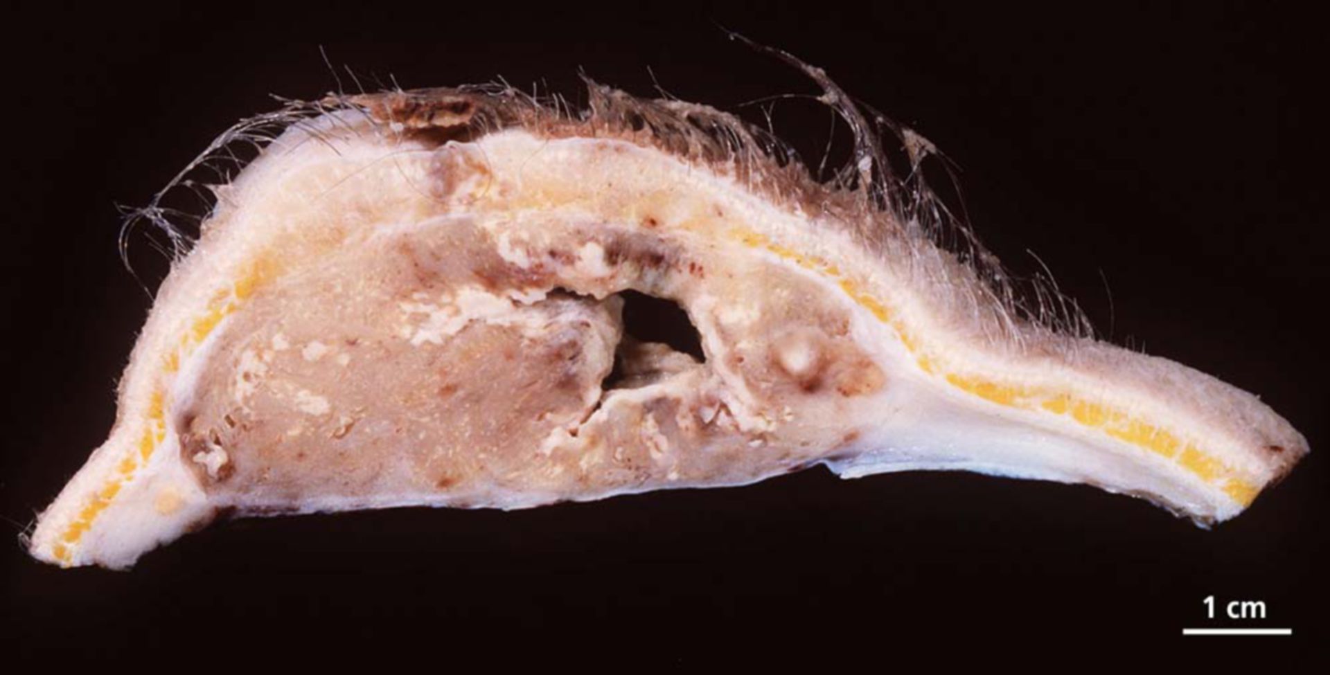
Cutis/ subcutis, 14 x 13 x 5 cm. Central tumor, around 2 cm thick, superficial ulcerating, 13 cm of size. Light brown tumor tissue with cystic necrosis.
Additional finding: Histological large infiltrations of a malign tumor, consisting of irregular vascular structures, laid out with pleomorph endothelial cells. In between are solid areas with spinocellular polymorph cells with large hyperchromatic nuclei. Multiple, partly atypical mitosis can be found. Large, partly landscape-shaped necrosis can be found as well. Immunohistochemically the tumor cells are positive for CD 31.
Clinical presentation: Metastasized angiosarcoma of the scalp.
Source: © PathoPic