The penis, together with the scrotum, forms the external male genital organs.
The penis can be subdivided anatomically into 3 sections:
The root of the penis is the proximal portion, attaching the penis to the bony pelvis. This attachment is supported by ligaments and pelvic floor muscles, particularly the bulbospongiosus and ischiocavernosus muscles. These muscles help maintain an erection by compressing veins to restrict venous outflow and assist with ejaculation through rhythmic contractions. Two notable ligaments are:
At the base of the penis, the two diverging crura of the penis and the bulb of the penis (an enlargement of the spongy tissue surrounding the urethra) are located.
The body of the penis makes up the majority of the organ's length. In cross-section, the body consists of three cylindrical structures of erectile tissue:
The two corpora cavernosa are separated by a thin connective tissue layer, called the septum penis. On the ventral side of the penis, a midline fusion called the penile raphe is visible.
The glans forms the distal portion of the penis, marking the transition from the body of the penis. This transition is outlined by a groove called the coronal sulcus. The glans contains a continuation of the spongy erectile tissue (corpus spongiosum glandis), giving it its characteristic rounded shape. The external opening of the urethra is located at the tip of the glans.
The skin covering the body of the penis is thin and highly mobile, forming a fold (the foreskin, or prepuce) that can retract to expose the glans. A small band of tissue, the frenulum, connects the underside of the glans to the prepuce, preventing the foreskin from retracting too far. Sebaceous glands located near the frenulum secrete lubricating substances.
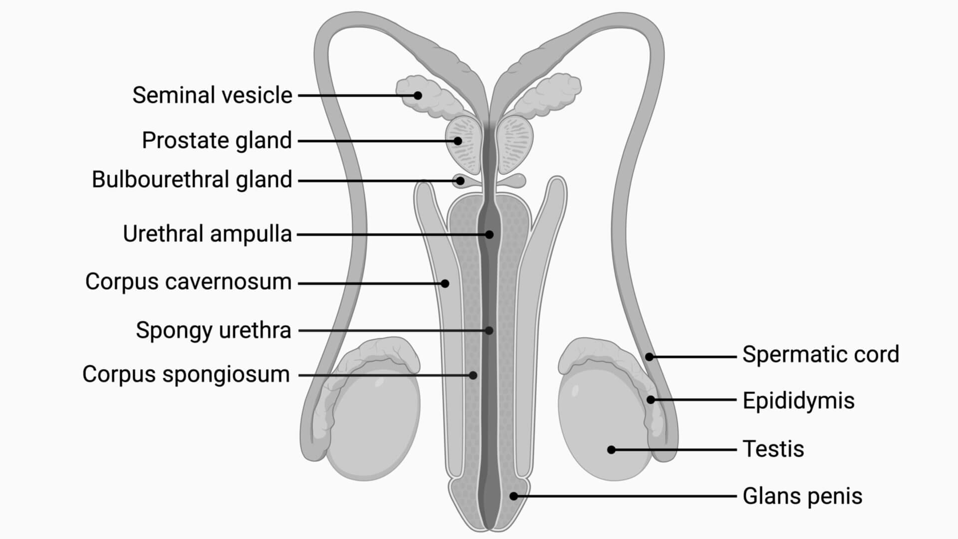
Author: Joshua Soeder, DocCheck, created with BioRender.com; adapted from " Male Reproductive System Anatomy" licensed under CC BY-NC-SA 3.0